Foreign Body Granuloma, Secondary to Retained Toothbrush Head
By Pir Abdul Ahad Aziz Qureshi1, Hafeez Ur Rehman Junejo1, Muhammad Talha Yaseen Kaimkhani2Affiliations
doi: 10.29271/jcpsp.2021.11.1378
Sir,
A 5-year boy presented with complaints of dysphagia and left-sided neck swelling for seven days. On examination, the swelling was hard in consistency and tender. The oral cavity was unremarkable. A smooth soft tissue mass with hyperemic mucosa was seen in the left oropharyngeal wall, extending up to the hypopharynx, causing luminal narrowing. There were no signs of bleeding, discharge or injury. CT scan revealed a large soft tissue mass in the left pharyngeal space extending into the upper visceral space with metal-like density within it. The mass was causing significant narrowing of the oropharynx and hypopharynx with obliteration of the left pyriform sinus and parapharyngeal space (Figure 1). 3-D images of the mass showed structure resembling toothbrush head (Figure 2). At this point, further history about the foreign body ingestion was asked from the patient’s parents, who revealed a vague history of toothbrush head ingestion by the child a couple of years back, followed by bleeding from mouth and dysphagia. He was treated with pain killers and antibiotics at a rural health facility for oral bleeding and dysphagia. The patient later remained asymptomatic until the recent presentation. Based on the clinical history and imaging findings, the diagnosis of foreign body granuloma was made. The patient underwent surgical removal of the foreign body under general anaesthesia. The patient is now asymptomatic at six weeks postoperative follow-up.
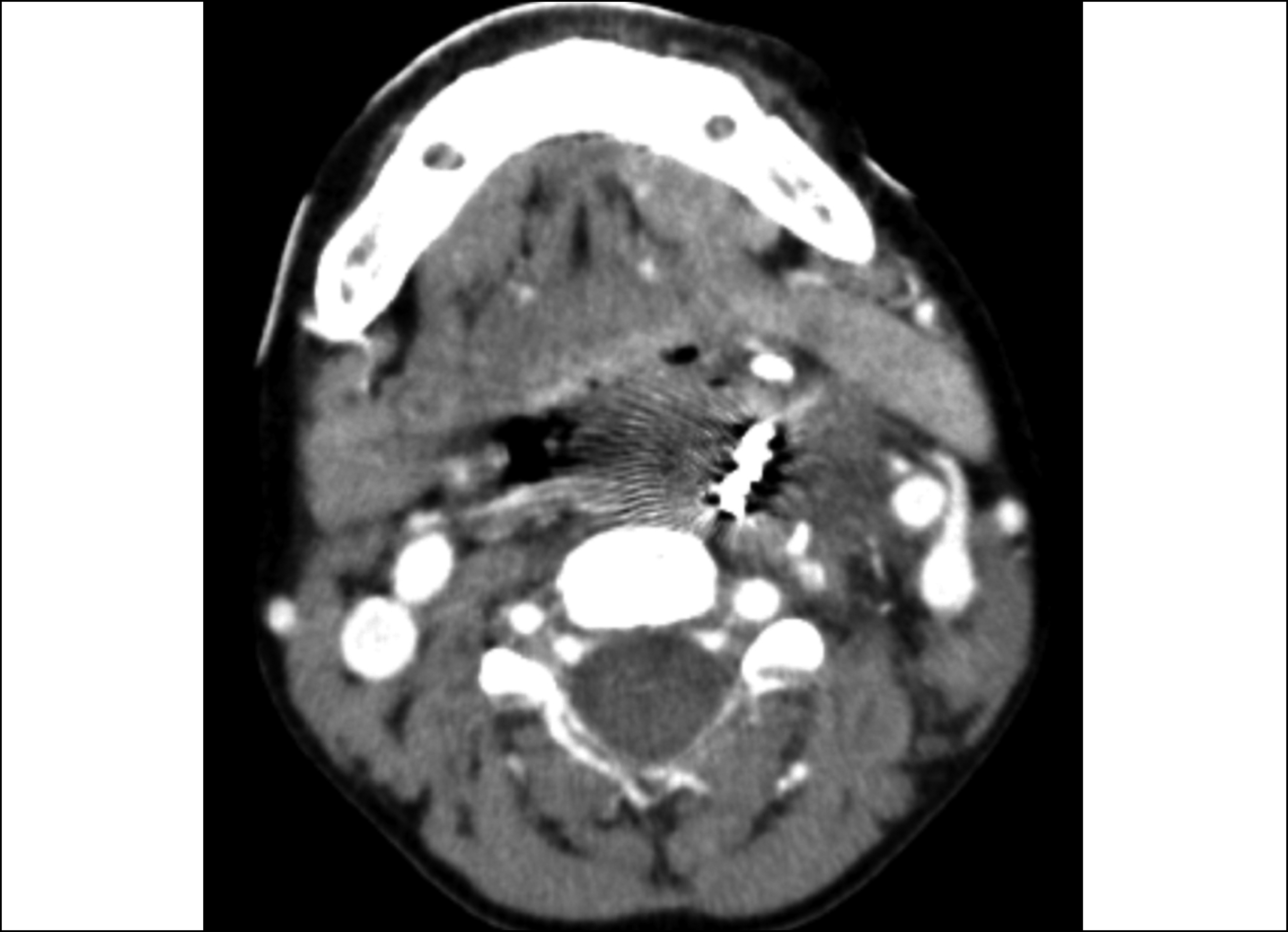 Figure 1: Contrast enhanced CT scan (axial view) showing large soft tissue mass in the left pharyngeal space with metal-like density seen within it.
Foreign body ingestion can occur in any age group; however, it is more common in infants and toddlers who tend to put everything in their mouth, which may become a medical/surgical emergency.1 The type of ingested foreign body varies greatly with the age groups: coins, buttons, button batteries, peas, grains, etc. being most common in infants and toddlers; and dentures, metallic pins, meat bones being most common in the adults.2 The body reacts to the retained foreign body by forming a tissue reaction around it, which is termed as foreign body granuloma.3,4 Foreign body granulomas are widely classified into two main groups, i.e., iatrogenic, as a result of retained surgical material (also called gossypiboma) and secondary to the penetrating injury.5
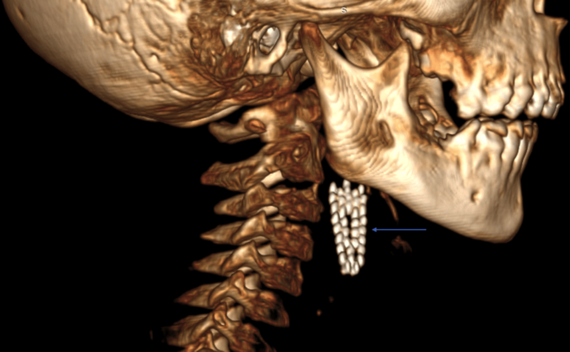 Figure 2: 3D image of the mass showing structure resembling tooth brush head (blue arrow).
Foreign body granuloma, secondary to retained toothbrush head, is very unusual and prompts quick medical attention.1 Careful correlation with clinical history and interpretation of images is required to make the correct diagnosis.
PATIENT’S CONSENT:
This case was granted waiver of informed consent by the Institutional Review Board.
CONFLICT OF INTEREST:
The authors declared no conflict of interest.
AUTHORS’ CONTRIBUTION:
PAAAQ: Wrote the manuscript.
HRJ: Performed the literature review.
MTYK: Collected the pictures and gathered important clinical information.
REFERENCES
- Farahnak MR, Araghi S, Nikakhlagh S, Saki N. Toothbrush: A report of an unusual foreign body. Iran J Otorhinolaryngol 2015; 27(80):247-9.
- Sharma RC, Dogra SS, Mahajan VK. Oro-pharyngo-laryngeal foreign bodies: Some interesting cases. Indian J Otolaryngol Head Neck Surg 2012; 64(2):197- 200. doi: 10.1007/s12070-011-0473-6.
- Ando A, Hatori M, Hagiwara Y, Isefuku S, Itoi E. Imaging features of foreign body granuloma in the lower extremities mimicking a soft tissue neoplasm. Upsala J Med Sci 2009; 114(1):46-51. doi:10.1080/0300973080 2602455.
- Sandhu GS, Parkash M, Bhatia V. Pseudotumor of hand foreign body granuloma. Indian J Musculoskelet Radiol 2019; 1(1):72-4. doi: 10.25259/IJMSR_2_2019.
- Perigela HC, Reddy K, Bangi VP, Janjala N. Foreign body granuloma mimicking a soft-tissue tumor. J NTR Univ Health Sci 2014; 3:143-5. doi: 10.4103/2277-8632.134899.