Relationship between Chronological Age and Dental Age Using Third Molar Calcification in the Pakistani Population
By Muhammad Bilal Bashir1, Syed Jaffar Abbas Zaidi2, Madiha Anwar3, Rafia Ruaaz1, Haifa S. Baqai4, Qaiser Ali Baig5
Affiliations
doi: 10.29271/jcpsp.2023.01.15ABSTRACT
Objective: To ascertain the relationship between dental age and chronological age in patients attending dental OPDs using third molar calcification stages.
Study Design: A cross-sectional analytical study.
Place and Duration of Study: Dental OPDs of Dow University of Health Sciences, Karachi, Pakistan, from November 2021 to April 2022.
Methodology: Dental Orthopantomograms (OPG) of 385 patients, aged 12–28 years attending the dental OPDs, were obtained during the study period. Third molar calcification stages were evaluated using the method proposed by Kohler et al. based on ten stages of tooth formation. The inter-examiner agreement was tested by adding another examiner to 100 dental OPGs after 30 days of reviewing them by one examiner. A simple linear regression model was applied between age and stage of tooth growth.
Results: A total of 121 males (31.4%) and 264 (68.6%) females were included in this study. Intra-agreement and inter-agreement were excellent (˃0.90). A total of 55 (14.28%) had half of the root completed, followed by 49 (12.72%) who had initiation of root formation and 1/3rd root completion. The left mandibular third molars were found to be more statistically significant with Kohler's stages of development in both genders.
Conclusion: Developing third molars was significantly related to chronological age and provided the most accurate age calculation based on all tooth measurements and ratios of tooth measurements. Third molar calcification stages can be used accurately to predict age in Pakistani adolescents.
Key Words: Dental age estimation, Chronological age, Dental radiographs, Orthopantomograph, Third molar development.
INTRODUCTION
Dentition in a person's oral cavity can best determine their age in the early childhood period; teeth aid in accurate and precise age estimation.1 However, this precision declines with an individual's advancing age. Furthermore, the process of teeth development can be affected by specific intrinsic and extrinsic factors such as genetic malformations, hormonal imbalances, nutritional deficiencies, and unfavourable climatic and environmental hazards.2
Furthermore, numerous studies have shown the effectiveness of developing dentition as a consistent and reliable age indicator tool.3-5
Dental age assessment can be performed by visually examining the erupted teeth through dental radiographs, which is the most helpful technique in addition to other reported methods.6 Accurate method of estimating the age from the development of dentition is when individual growth is rapid and many teeth are in the process of development.7 But once the teeth have completed their development, after age fourteen, assessing chronological age becomes more difficult.8 Later, only the developing third molars help estimate a person’s age.9
Numerous methods and techniques have been reported in chronological age estimation in juveniles and adults. These include radiographical analysis of the hand and wrist, the medial clavicular epiphyseal cartilage, and third-molar developmental observations.10-12
The evaluation of the chronological age of third molar calcification is recommended due to the lack of other reliable bone and cartilage biological markers during late puberty.12 All other permanent dentition complete their development at puberty as the root apices are closed.13 Kohler stages of tooth calcification were formulated for permanent molars with no variation for wisdom teeth.14 Among all the teeth in the dentition, third molars are the most inconstant and atypical in terms of anatomy,15 eruption age,6,9 and agenesis.9 In late puberty, age assessment becomes less reliable with biological bone and cartilage markers, whereas it becomes more reliable with third molar calcification. Third molar development befalls earlier in males than in females.1,9,17
In the permanent dentition stage, which correlates with puberty, wisdom teeth mineralisation stages are reliable indicators of age. The stage of development and calcification of the third molar has a linear relationship with the age of the patients regardless of gender.9
There are ethnic differences in estimating chronological and dental age.4 There is a paucity of local data to establish an association between chronological and dental age. There is a need to fill this gap in the literature and assist in compiling a national database for estimating dental age and legal age for forensic odontology and medico-legal purposes. The objective of this study was to find the relationship between dental age and chronological age in patients attending dental OPDs using third molar calcification stages.
METHODOLOGY
This cross-sectional study was conducted from November 2021 to April, 2022 on 385 patients aged 12-28 years attending dental OPDs at Dow University of Health Sciences. Dental OPG X-rays of these patients were taken after informed consent. The third molar calcification stages were evaluated using the method proposed by Kohler et al. based on the ten stages of tooth formation. The study was approved by the Institutional Review Board of Dow University of Health Sciences (Ref: IRB-2236/DUHS/Approval/2021/570 dated 8th November 2021).
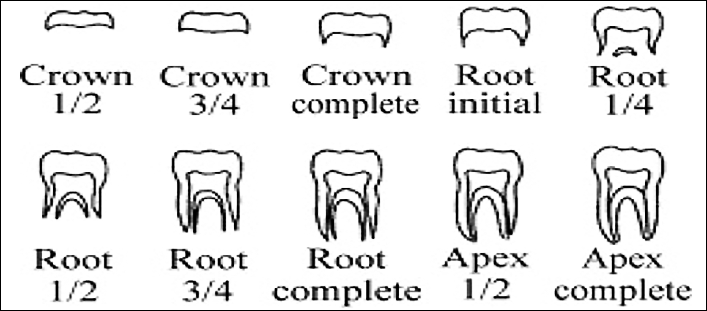 Figure 1: Developmental stages according to the 10-point scoring system developed by Gleiser and Hunt and modified by Kohler.
The evaluation of third molar calcification was based on Kohler et al. This evaluation was based on ten stages of tooth formation.14 After calculating age from the third molar calcification stages, it was correlated with patients' chronological age, similar to previous regional studies.17 The first four stages (1–4) show crown formation from the initiation of cusp calcification until the completed crown, the following four (5–8) stages show root formation starting from the initiation of radicular bifurcation to root completion, and the last two (9-10) stages consist of apical formation.14 The developmental stages of the third molar from Dental OPGs were matched with the diagrammatic representation of Kohler's stages as shown in Figure 1.14
The sample size for this study was determined using the WHO sample size calculator by estimating a population proportion with specified absolute precision, maintaining a 95% confidence interval, 0.005 expected population proportion, 0.20 precision, and 5% margin of error, resulting in a total of n=385.
Any developmental tooth anomaly affecting the third molar, missing mandibular third molars, and image deformity affecting the third molars were excluded from this study. Inclusion criteria included patients between the ages of 12 till 28 years, mandibular third molars present, and patients undergoing dental treatment. Research participants had no risks since dental X-rays were taken for routine dental treatment. No additional X-rays were obtained for this study. Data were stored on Google Drive and Google Cloud and were accessible to the authors. The patient's details, such as their names and contact details, were confidential and not shared with anyone. To mark the development of the third molar, radiographic records were matched with the diagrammatic representation.
Statistical test was performed using SPSS version 20. The categorical variables were expressed as counts and percentages, and continuous variables were expressed as mean and standard deviations. The Chi-square test was applied to assess the association between different stages of tooth eruption and gender. The Chi-square test was applied, considering a p-value of 0.05 or less as statistically significant. An inter-examiner agreement was tested by adding another examiner to evaluate 100 dental OPGs after 30 days of reviewing them by one examiner. Intraclass correlation coefficient (ICC) was used to test intra-agreement between the observers. ICC was calculated on a 2-way mixed model. The variables were the measurements of the length of crown and roots on dental OPGs corresponding to the developmental stages based on the 10-point scoring system developed by Gleiser and Hunt and modified by Kohler as shown in Figure 1. ImageJR software (https://imagej. nih.gov/ij/) was used to generate the parameters of the crown and roots through measuring the distance from the cusp tip to the desired landmark using the straight-line feature of ImageJ as shown in Figure 2. Once the yellow line was drawn, it was measured in pixels and different results corresponding to various aspects of the third molar were measured by the first observer and then the second observer. ImageJR is a reliable freeware for accurately measuring distances on radiographs and calculating areas on microscopic slides. The intra-agreement and inter-agreement based on the intraclass correlation coefficient (ICC) were excellent, with a value of ˃0.90 and a Cronbach's alpha value of 1.0. ICC can take a value from 0 to 1, with 0 indicating no agreement and 1 indicating perfect agreement.18 A simple linear regression model was applied between age and stage of tooth development growth.
Table I: The association between different stages of tooth eruption on both sides of the mandible with gender.|
Stages of Eruption |
Left Mandibular 3rd Molar (N=385) |
Right Mandibular 3rd Molar (N=385) |
||
|
Males N (%) |
Females N (%) |
Males N (%) |
Females N (%) |
|
|
Stage 1 – Crown Half made |
02 (1.70) |
04 (1.5) |
03 (2.5) |
05 (1.9) |
|
Stage 2 – Crown-3/4 made |
06 (5.0) |
02 (0.8) |
06 (5.0) |
07 (2.7) |
|
Stage 3–Crown completion |
04 (3.3) |
18 (6.8) |
3 (2.5) |
13 (4.9) |
|
Stage 4 - Root Initiation |
20 (16.5) |
29 (11.0) |
19 (15.7) |
30 (11.4) |
|
Stage 5 – Root ¼ made |
17 (14.0) |
32 (12.1) |
18 (14.9) |
31 (11.7) |
|
Stage 6 – Root ½ made |
16 (13.2) |
38 (14.4) |
16 (13.22) |
39 (14.8) |
|
Stage 7 – Root ¾ made |
0 (0) |
23 (8.7) |
00 (0) |
25 (9.5) |
|
Stage 8 – Root Completion |
13 (10.7) |
33 (12.5) |
13 (10.7) |
37 (14.0) |
|
Stage 9 – Apex half made |
16 (13.2) |
38 (14.3) |
16 (13.2) |
30 (11.4) |
|
Stage 10 – Apex Complete |
27 (22.3) |
47 (17.8) |
27 (22.3) |
47 (17.8) |
|
Total |
121 |
264 |
121 |
264 |
|
p-value * |
0.006 |
0.036 |
||
|
* Chi-Square Test was applied, considering a p-value of 0.05 or less as statistically significant. |
||||
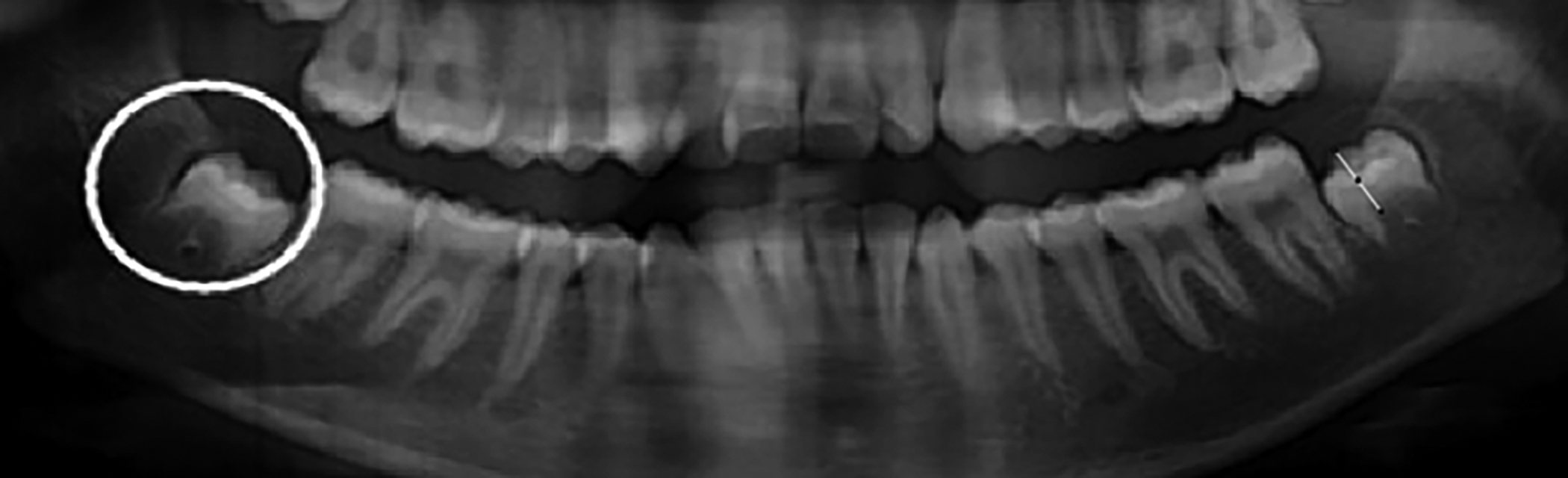 Figure 2: Measuring crown and root parameters through ImageJ for intra-agreement for ICC.
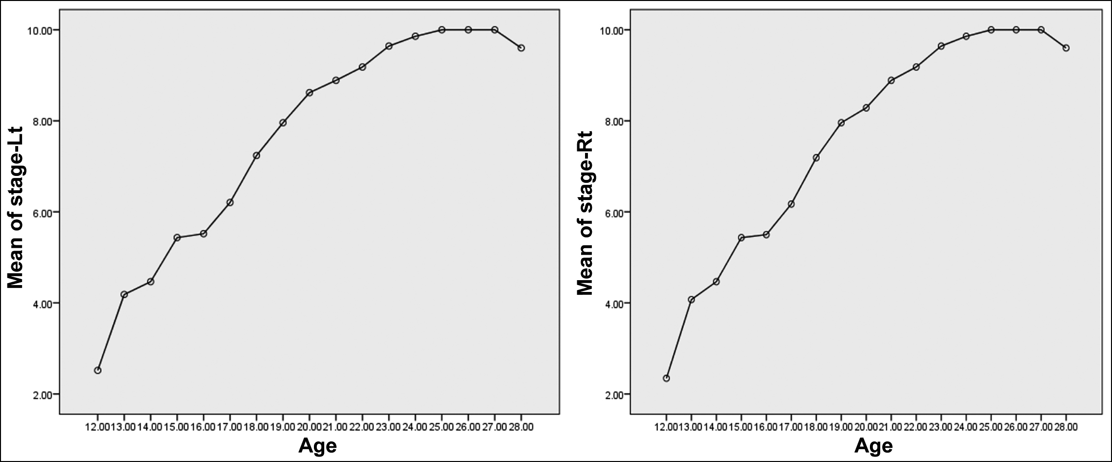 Figure 3: Distribution of different stages of the eruption of the right and left side of the mandible according to age.
RESULTS
In this study, there were 385 participants with 121 males (31.4%) and 264 (68.6%) females. The minimum age recorded was 12 years, and the maximum age was 28 years, with a mean age of 17.9 years. The mandibular third molars were visually present bilaterally on all the 385 participants' radiographs with all their landmarks visible. Records were sub-classified by gender and age for both right and left mandibular hemi-arches. There were 27 males (7.01%) and 47 females (12.21%) that had fully developed third molars. In this study, 55 (14.28%) had half root completed, followed by 49 (12.72%) who had initiation of root formation and 1/3rd root completion, as shown in Figure 3. The distribution of different stages of the eruption of the right and left side of the mandible according to age is shown in Figure 3. The association for different stages of tooth eruption on both sides of the mandible was assessed with gender. On the left side, teeth eruption was found to be more statistically significant with different stages in both genders (p = 0.006) compared to the mandibular right side (p = 0.036), as shown in Table I.
Simple linear regression was used to determine whether age significantly predicted tooth development. The regression was statistically significant (R2 = 0.702, F (1, 383) = 901.3, p = 0.0001) for the right side of the maxillary and mandibular arch and also for the left side of the maxillary and mandibular arch (R2 = 0.711, F (1, 383) = 943.365, p = 0.0001). It was found that age significantly predicted right and left arches in terms of tooth development (β = 0.529 (left side), 0.533 (right side) p = 0.0001).
DISCUSSION
The evaluation of chronological age in the development stages of the third molar is recommended due to the lack of other reliable biological markers during late puberty.16 Permanent dentition, except for the third molar, has completed its development by puberty in adolescents.13 Precision and accuracy are improved when many teeth develop when calculating chronological age. In this study, the Kohler stages of developing teeth were used for permanent molars with no variation for wisdom teeth. Third molars are the most inconstant teeth in the entire dentition based on their anatomy15, eruption age,6 and agenesis.7 The third molar follicle or crypt formation is usually observable from seven to nine years in both the mandible and the maxilla. The diagnosis of agenesis can be conclusively determined if there is no dental follicle or radiolucent bud at the age of 14 years.
Third molar development befalls earlier in males than in females. This conclusion endorses the trend stated by Moorrees et al.,16 Grill et al.10,11, and Kullman et al.1 In a Belgian study, the mean age at each developmental stage was lower for males than for females, indicating a slightly earlier development in males compared to females.19 There is also a trend for earlier third molar development in the maxilla than in the mandible, as endorsed by various population-based studies.17,20,21
The age approximation of tooth development lacks reliability after reaching stage 10, as apices are closed by that age. Stages 8-10, which represent late root and apex formation, are predominantly associated with individuals at or above 18 years of age. The presence of these stages on radiographs provides valuable information about the legal age of the individual. Inferences on the legal age should only be made using multiple parameters and the development of other teeth due to the broad variability of third molar development and its frequent congenital absence.
In this study, the mean age for stage 10 was 17.95 years. However, the mean may be lower if the sample age range is restricted to patients younger than 24 years. A Malay study showed that females at the age of 18 years reached the third molar stage 7 while their male counterparts were in stage 8 at the same age.22 For males and females in the Russian population, stage 8 appears at 18 years.23 Therefore, the authors suggest that the maximum age limit of patients in further research should not be greater than 22 years. This apparent variance between different populations may be attributed to methodological differences and the exclusion of third molars. Another aspect attributed to the variability in estimating dental age is differences in viewing conditions, as the radiographic interpretation depends on them.24
Furthermore, CBCT is more reliable and provides a comprehensive view of root and crown formation than a two-dimensional OPG. The prohibitive cost of CBCT precludes its routine use, especially for dental screening in third-world countries. The third molar calcification stage is one of the tools that can be used to measure age when development is near completion during puberty when the third molar is the only viable dental indicator.
The developmental stage of the third molar has a linear relation to the age of the patients, whether female or male. Statistical analysis shows a stronger correlation between male and female patients in this study. These results concord with an OPG-based study conducted on Turkish patients ranging from 4-20 years.15 Table I shows a strong correlation between age and third molar development for both males and females.
Furthermore, the eruption of the third molar on the left side of the mandible was found to be more statistically significant with various stages in all genders compared to the right mandibular side. This left-right symmetry of the mandibular third molars has been noted in a Belgian OPG-based study of individuals ranging from 15.7 and 23.3 years.19 The left mandibular teeth have also been shown to agree better with the stages of tooth development than their maxillary counterparts in a study conducted in India.25 This can be an important population-specific finding attributed to the Pakistani population.
However, age estimation can be more challenging when individuals present with third molars in the late stages of root formation. Nonetheless, third molar staging can be used as an adjunctive tool to make inferences on the legal age. Congenital absence of third molars, known as agenesis, is a barrier to reliable age estimation. However, when third molars are present, legal can be reliably predicted based on the developmental stages of the crown and root formation.
This study has practical implications in forensic odontology that involves estimating dental age on judicial request. Based on the findings of this study, third molars can serve as a useful developmental marker. The development stages of the third molars can predict age more reliably than other skeletal age calculation techniques, such as hand-wrist radiographs and cervical ribs radiographs.21 Using third molars along with the development stages of other teeth adds to the reliability of dental age estimation.
Further research should be aimed at developing a national database of dental radiographs and a policy of storing dental OPGs of patients by dentists and radiologists. These databases will help improve the dental and forensic age estimates based on the mineralisation of teeth.
CONCLUSION
Developing third molars was most strongly associated with age and provided the most accurate age calculation based on all tooth measurements and ratios of tooth measurements. Uniting the third molar recording with tooth measurement or ratios did not subsidize a clinical-related information gain for age prediction.
Therefore, the process of third molar staging and associated recording must be acclaimed over complex dimension measurements or ratio calculations of the third molar for age assessment.
ETHICAL APPROVAL:
Institutional Review Committee of Dow University of Health Sciences approved this study (Ref: IRB-2236/DUHS/Approval/2021/570 dated 8th November 2021). Ethical approvals were obtained prior to initiation of the research work.
PATIENTS’ CONSENT:
Written informed consent were obtained from patients to publish the data concerning this case.
COMPETING INTEREST:
The authors declared competing interest.
AUTHORS’ CONTRIBUTION:
MBB: Research design and manuscript writing.
SJAZ: Conceived this study and critically analysed the manuscript.
MA: Statistical analysis and manuscript writing.
RR: Analysed the data and critically reviewed the manuscript.
HSB: Data collection and manuscript writing.
QAB: Statistical analysis and manuscript writing.
All authors gave final approval and agree to be accountable for all aspects of the work.
REFERENCES
- Kullman L. Accuracy of two dental and one skeletal age estimation method in Swedish adolescents. Forensic Sci Int Oct 1995; 75(2-3):225-36. doi.org/10.1016/0379-0738 (95) 01792-5.
- Lewis AB, Garn SM. The relationship between tooth formation and other maturational factors. Angle Orthodontist 1960; 30(2):70-7 doi.org/10.1043/0003-3219 (1960)030% 3C0070: TRBTFA%3E2.0.CO;2.
- Kurita LM, Menezes AV, Casanova MS, Haiter-Neto F. Dental maturity as an indicator of chronological age: Radiographic assessment of dental age in a Brazilian population. J Applied Oral Science 2007; 15:99-104. doi.org/10.1590/ S1678-77572007000200005.
- AlQahtani S, Hector M, Liversidge H. Accuracy of dental age estimation charts: Schour and Massler, Ubelaker and the London Atlas. American J Physical Anthropol 2014; 154(1): 70-8. doi.org/10.1002/ajpa.22473.
- AlQahtani SJ, Hector MP, Liversidge HM. Brief communication: The London atlas of human tooth development and eruption. Am J Phys Anthropol 142(3):481-90. doi:10. 1002/ajpa.21258.
- Prieto JL, Barbería E, Ortega R, Magaña C. Evaluation of chronological age based on third molar development in the Spanish population. Int J Legal Medicine 2005; 119(6): 349-354. doi.org/10.1007/s00414-005-0530-3.
- Hagg U, Matsson L. Dental maturity as an indicator of chronological age: the accuracy and precision of three methods. European J Orthodontics 1985; 7(1):25-34. doi.org/10.1093/ejo/7.1.25.
- Maber M, Liversidge HM, Hector MP. Accuracy of age estimation of radiographic methods using developing teeth. Forensic Sci Int May; 159 (Suppl) 1:S68-73. doi:10.1016/j. forsciint.2006.02.019.
- Thorson J, Hägg U. The accuracy and precision of the third mandibular molar as an indicator of chronological age. Swedish Dental J 1991; 15(1):15-22. doi.org/10.1093/ ejo/ 7.1.25.
- Grill F, Franke J. The llizarov distractor for the correction of relapsed or neglected clubfoot. J Bone and joint Surg 1959; 69(4):593-7. doi: 10.1302/0301-620X.69B4.3611163.
- Kreitner K-F, Schweden F, Riepert T, Nafe B, Thelen M. Bone age determination based on the study of the medial extremity of the clavicle. European Radiol 1998; 8(7): 1116-1122. doi.org/10.1007/s003300050518.
- Olze A, Schmeling A, Taniguchi M, et al. Forensic age estimation in living subjects: the ethnic factor in wisdom tooth mineralization. Int J Legal Med 2004; 118(3):170-3. doi.org/10.1007/s00414-004-0434-7.
- Arany S, Iino M, Yoshioka N. Radiographic survey of third molar development in relation to chronological age among Japanese juveniles. J Forensic Science 2004; 49(3): JFS2003372-5. doi.org/10.1520/JFS2003372.
- Kohler S, Schmelzle R, Loitz C, Puschel K. Development of wisdom teeth as a criterion of age determination. Ann Anat 1994; 176(4):339-45. doi.org/10.1016/s0940-9602(11) 80513-3.
- Orhan K, Ozer L, Orhan A, Dogan S, Paksoy C. Radiographic evaluation of third molar development in relation to chronological age among Turkish children and youth. Forensic Science Int 2007; 165(1):46-51. doi.org/10.1016/j. forsciint.2006.02.046.
- Moorrees CF, Fanning EA, Hunt EE. Age variation of formation stages for ten permanent teeth. J Dental Res 1963; 42(6):1490-1502. doi.org/10.1177/00220345630420062 701.
- Bagherpour A, Anbiaee N, Partovi P, Golestani S, Afzalinasab S. Dental age assessment of young Iranian adults using third molars: A multivariate regression study. J Forensic Leg Med Oct; 19(7):407-12. doi:10.1016/j.jflm. 2012.04.009.
- Ranganathan P, Pramesh C, Aggarwal R. Common pitfalls in statistical analysis: Measures of agreement. Perspectives Clinical Research 2017; 8(4):187. doi: 10.4103/picr.PICR_ 123_17.
- Gunst K, Mesotten K, Carbonez A, Willems G. Third molar root development in relation to chronological age: A large sample sized retrospective study. Forensic Science Int 2003; 136(1-3):52-57. doi.org/10.1016/S0379-0738(03) 00263-9.
- Karatas OH, Oztürk F, Dedeoglu N, Çolak C, Altun O. Radiographic evaluation of third-molar development in relation to the chronological age of Turkish children in the southwest Eastern Anatolia region. Forensic Sci Int 2013; 232(1-3):238.e1-5. doi:10.1016/j.forsciint.2013.07.023.
- Thevissen P, Alqerban A, Asaumi J. Human dental age estimation using third molar developmental stages: Accuracy of age predictions not using country specific information. Forensic Science Int 2010; 201(1-3):106-111. doi.org/ 10.1007/s00414-009-0329-8.
- Yusof MYPM, Cauwels R, Martens L. Stages in third molar development and eruption to estimate the 18-year threshold Malay juvenile. Archives Oral Biology 2015; 60(10): 1571-1576. doi.org/10.1016/j.archoralbio.2015.07. 017.
- Franco RPAV, Franco A, Turkina A. Radiographic assessment of third molar development in a Russian population to determine the age of majority. Archives of Oral Biology 2021; 125:105102. doi.org/10.1016/j. archoralbio.2021. 105102.
- Moshfeghi M, Shahbazian M, Sajadi SS, Sajadi S, Ansari H. Effects of different viewing conditions on radiographic interpretation. J Dentistry (Tehran, Iran) 2015; 12(11):853. PMID: 27507997; PMCID: PMC4977410.
- Raj LM, Jude J, Anison JJ, Srikant N, Krishna PS, Shankar K. Age estimation with third molars using orthopantomograph (OPG): An Indian scenario. Indian J Forensic Odontology Volume 2015; 8(3-4). doi.10.4103/ijfo.ijfo_16_18.