Radiological Insight into the Mermaid Syndrome: An Exploration of a Rare Anomaly
By Kanwal Laique, Zanobia Saira Waseem, Kainat NaseemAffiliations
doi: 10.29271/jcpsp.2024.03.377Sir,
Mermaid syndrome is an extremely rare lethal congenital malformation which is characterised by complete or incomplete fusion of both lower limbs. It is associated with absent urinary bladder, external genitalia, imperforate anus, renal agenesis, etc.1
Here, we report a case of a 1-day neonate weighing 1.6 kg who presented with respiratory distress since birth. The antenatal history showed prolonged labor and preterm spontaneous vaginal delivery at 34 gestational weeks in a hospital. The baby did not cry immediately at birth.
On clinical neonatal examination, the baby had no anal and urethral openings. Both lower limbs were fused. The body was fixed in a flexion contracture position (Figure 1). The airway was clear and work of breathing was increased. Moro's reflex was weak. The rest of the neonatal examinations were unremarkable. No antenatal ultrasound was done due to unsupervised pregnancy.
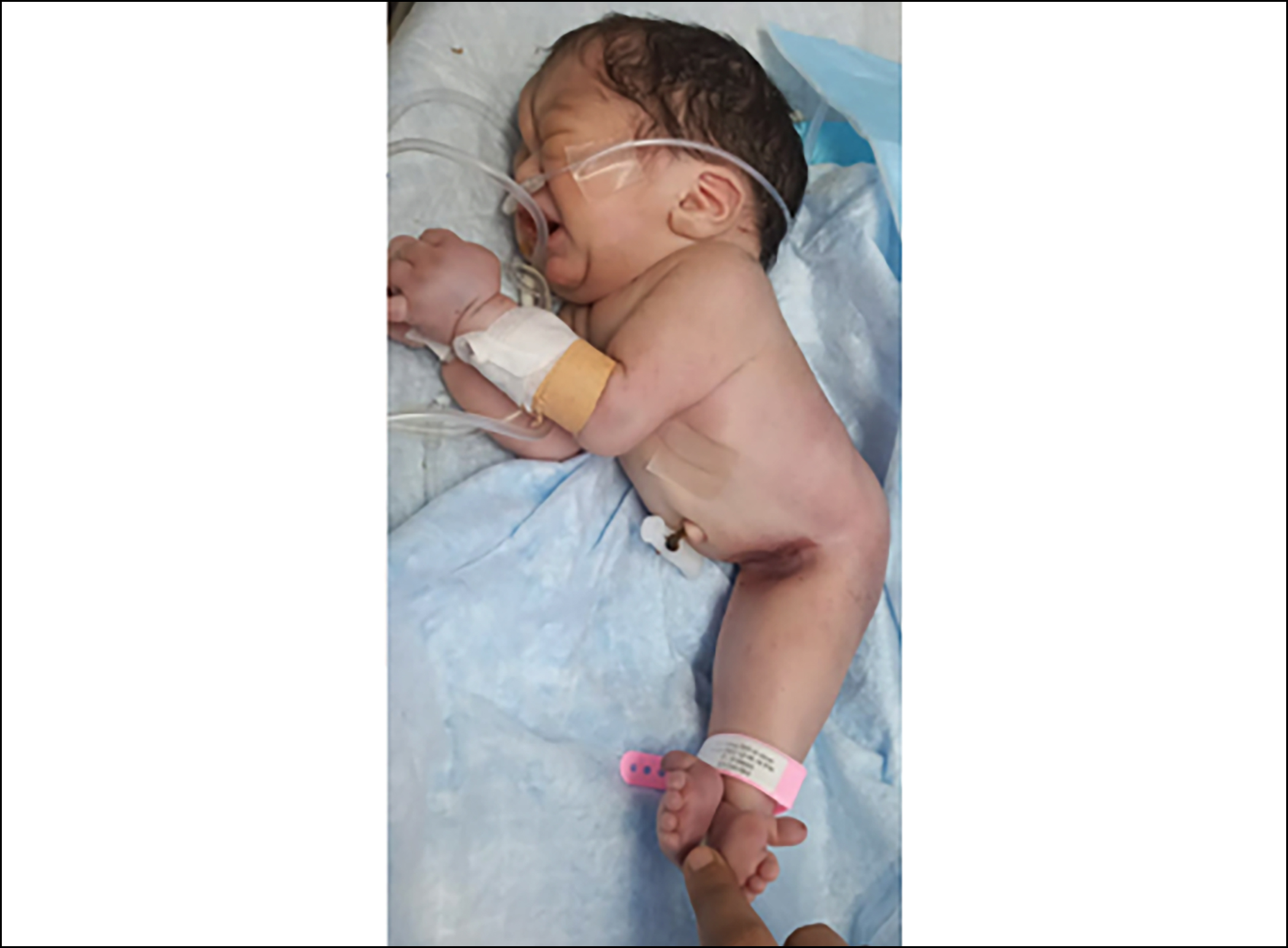 Figure 1: Clinical photograph of the patient.
After birth, he underwent a baby-gram which demonstrated absent sacrum, pubis and ischium, one soft tissue shaft of the lower limb with two separate femurs, tibias and fibulas (Figure 2). His feet were also fused and facing anteriorly but the metatarsals and phalanges were grossly normal. Post-natal ultrasound abdomen showed bilateral small-sized kidneys with multiple simple renal cortical cysts replacing whole parenchyma. Additionally, two small structures resembling testicles were also noted.
Mermaid syndrome is a severe form of caudal regression syndrome. The prevalence of this malformation ranges from 0.1 to 1% with male-to-female ratio of 2.7:1.2
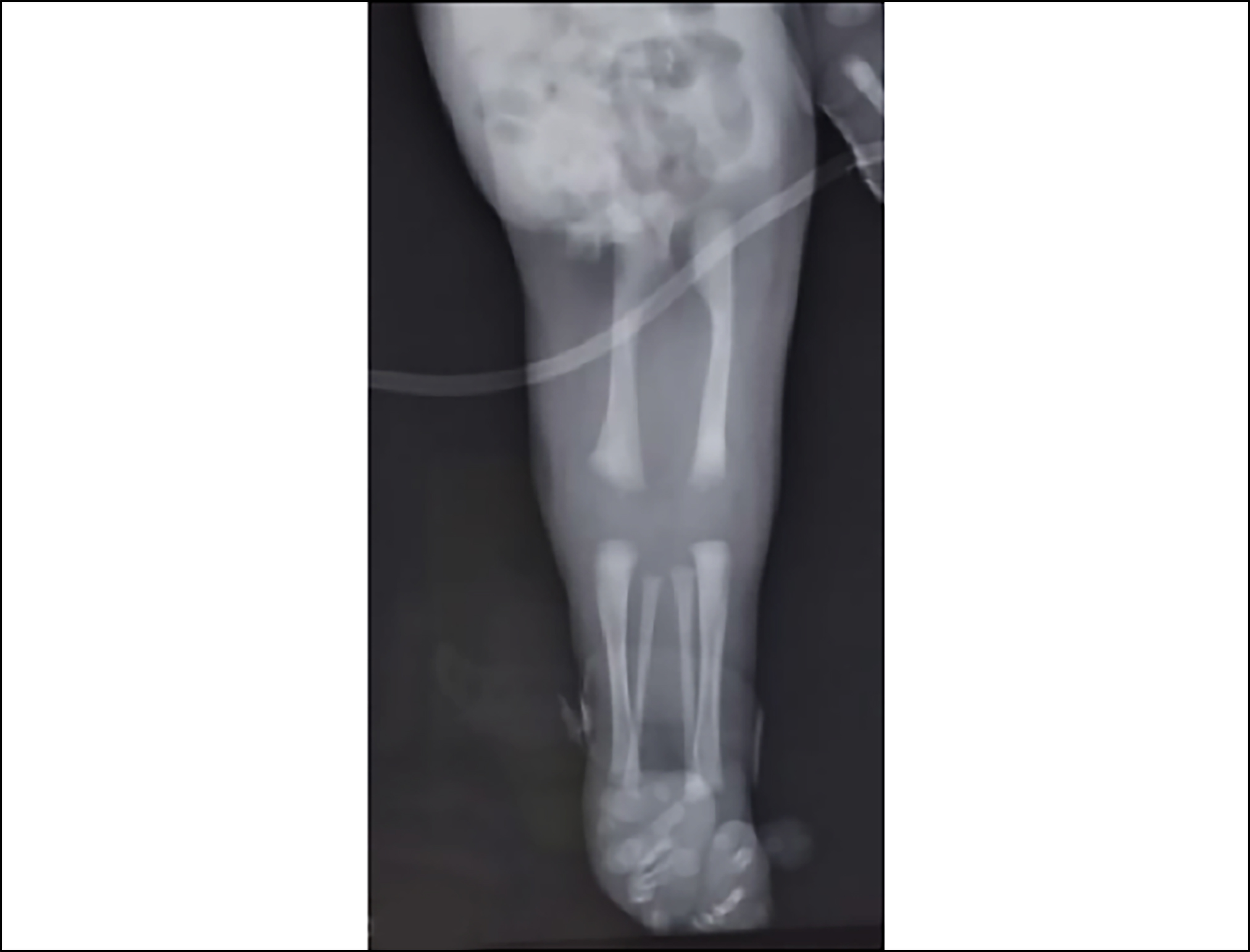 Figure 2: X-ray of the fused lower limbs of the neonate.
The exact aetiology is unknown. However, it is postulated that it is a combination of genetic predisposition and environmental trigger factors. Two main proposed pathogenic hypotheses are: the vascular steal hypothesis and the defective blastogenesis hypothesis. Tobacco use, heavy metal exposure, maternal diabetes, and retinoids are possible environmental causative agents. However, in the present case, no such environmental factor was found.
Approximately 300 cases have been reported in the literature, and most of the cases were diagnosed after birth. Mermaid syndrome can be diagnosed as early as 13 weeks of pregnancy. Antenatal grey and Doppler ultrasound are of utmost importance not only for the presence of a single umbilical artery and persistent vitelline duct which are the pathognomic features of Mermaid syndrome but also for oligohydramnios, renal agenesis, and absent bladder.3
Conventional radiology also helps in diagnosing the extent of the anomaly. Stocker and Heifetz's classification of Mermaid syndrome is commonly accepted. It has seven classifications based on the presence or absence of femurs, tibias and fibulas.4 However, postmortem examination can determine the nature and severity of the disease.
It has a very poor prognosis with a rare survival beyond five days. Post-natal surgical management of Mermaid syndrome is difficult, particularly when present with genitourinary and gastrointestinal malformations, with unpredictable results. Hence, genetic counselling and antenatal folate supplement intake should be offered. Pre-natal diagnosis should also be emphasised for timely optimal management that would be consequently less demanding from both parental psychological and healthcare cost points of view.5
PATIENTS’ CONSENT:
The consent was taken from the parents of the patient for publication of the case report including CT images.
COMPETING INTEREST:
The authors declared no competing interest.
AUTHORS’ CONTRIBUTION:
KL: Drafting, editing, review, and final approval.
ZSW: Acquisition of all medical details, literature search, and manuscript review.
KM: Drafting and manuscript review.
All authors approved the final version of the manuscript for publication.
REFERENCES
- Yaşar MZ, Yusuf AA, Hassan FM, Ali AA, Roble MA. Mermaid syndrome: A case report in Somalia. Ann Med Surg 2022; 76:103533. doi: 10.1016/j.amsu.2022.103533.
- Dharmraj M, Gaur S. Sirenomelia: A rare case of foetal congenital anomaly. J Clin Neonatol 2012; 1(4):221-3. doi: 10.4103/2249-4847.106006.
- Samal SK, Rathod S. Sirenomelia: The mermaid syndrome: Report of two cases. J Nat Sci Biol Med 2015; 6(1):264-6. doi: 10.4103/0976-9668.149227.
- Rani A, Sultan M. Antenatal diagnosis of sirenomelia, the mermaid syndrome with bilateral renal agenesis. Case Rep Clin Radiol 2023; 1(2):92-5. doi:10.25259/CRCR_25_2022.
- Fadhlaoui A, Khrouf M, Gaigi S, Zhioua F, Chaker A. The sirenomelia sequence: A case history. Clin Med Insights Case Rep 2010; 3: 41-9. doi: 10.4137/ccrep.s5347.