Métaizeau Closed Reduction Technique and Open Reduction Technique for the Surgical Treatment of Pediatric Radial Neck Fractures
By Murat Yilmaz, Mahmud Aydin, Melih MalkocAffiliations
doi: 10.29271/jcpsp.2022.05.613ABSTRACT
Objective: To compare the functional outcomes of pediatric radial neck fractures treated with percutaneous reduction using Kirschner (K) wire with the Métaizeau technique, and that with open reduction plus internal fixation with K-wire.
Study Design: Comparative descriptive study.
Place and Duration of Study: Haseki Training and Research Hospital Orthopedics and Traumatology Department, from December 2007 to December 2018.
Methodology: Children aged under 15 years, with radial neck fractures were inducted. The injury was classified according to Judet classification and the type IV was treated with either of the above technique. The inclusion criteria were a diagnosis of Judet type IV radial neck fracture and a minimum follow-up of 12 months. Exclusion criteria were patients with concomitant elbow fracture, follow-up for <12 months, failure to complete the Mayo Elbow Performance Score (MEPS) functional assessment, patients with missing data. Radiological results were evaluated in accordance with the Ursei Classification. MEPS was used to assess functional development.
Results: Forty-seven children (25 boys and 22 girls) aged (5–14 years with mean age of 8.57 ± 2.3 years were inducted. The surgical approach was the Métaizeau technique in 22 patients and open reduction technique in 25 patients. MEPS in the Métaizeau technique group was 95.2, with excellent results in 15 patients (68%), good results in 7 (31%), and fair or poor results in none of the patients. The mean MEPS in the open reduction / K-wire group was 88, with excellent, good, fair, and poor results in 9 (36%), 12 (48%), 4 (16%), and none of the patients, respectively.
Conclusion: Closed reduction using the Métaizeau technique with the elastic stable intramedullary nailing method satisfies all the criteria for minimally invasive bone surgery. This approach was forward to be considerably efficient, with excellent functional and esthetic outcomes and a low rate of complications if the indications and biomechanical principles are considered.
Key Words: Radial neck, Métaizeau technique, Judet classification.
INTRODUCTION
Radial neck fractures are the third most common type of elbow fracture in children.1 These fractures often occur in childhood around the age of 5 years, when the epiphyses begin to ossify.2 The mechanism of this fracture pattern typically occurs when the elbow is subjected to valgus loading while the elbow joint is extended.3
The displacement and angulation of the fracture provide the main guidance for choosing the treatment approach in pediatric radial neck fractures.2,4 These fractures are difficult to treat and can cause limited elbow motion; in addition, the management of radial neck fractures in children remains controversial.5 Although minor degrees of angulation can be acceptable for treatment with immobilisation alone, displaced fractures are rare and may require surgery.6
In the surgical treatment of displaced radial neck fractures, open reduction plus internal fixation with Kirschner wire (K-wire), percutaneous leverage reduction, and the Métaizeau technique and arthroscopically assisted reduction and pinning are currently used.7,8 There are studies comparing surgical treatments in the literature, but it has not been clearly stated which treatment method is superior.
The aim of this study was to compare the functional outcomes of pediatric radial neck fractures between treatment with percutaneous reduction using K-wire with the Métaizeau technique and open reduction plus internal fixation with the K-wire technique. The hypothesis of the study was that clinical outcomes would be better in the group of patients operated on with the Métaizeau closed reduction technique.
METHODOLOGY
This study was conducted retrospectively in accordance with the ethical standards of the SBU Haseki Training and Research Hospital Clinical Research Ethics Committee and the 1975 Declaration of Helsinki revised in 2013. Ethics committee approval was obtained (Decision No. 2020-27, 25/11/2020). All pediatric patients admitted to the emergency department and treated for radial neck fractures between December 2007 and December 2018 were screened using hospital digital records and patient files. The Judet classification was used for classifying the radial neck fractures (Table I).9 Closed reduction was performed in the patients by routinely applying the Israeli technique, and Judet type 3 and 4 fractures with unsuccessful closed reductions were referred for surgical treatment.10 The inclusion criteria were a diagnosis of Judet type IV radial neck fracture and a minimum follow-up of 12 months. Exclusion criteria were patients with concomitant elbow fracture, follow-up for <12 months, failure to complete the Mayo Elbow Performance Score (MEPS) functional assessment, and patients with missing data. All the surgeries were performed by two experienced pediatric surgeons. Depending on the preference of the treating surgeon, the operation was performed with one of the two different surgical techniques and follow-up. The surgical approach was percutaneous leverage reduction with the Métaizeau technique in 22 patients and open reduction plus internal fixation with the K-wire technique in 25 patients (Figures 1 and 2).
All the procedures were performed under general anesthesia, with tourniquet and fluoroscopy control. All the patients were given intravenous first-generation cephalosporin (1 g cefazolin) 1 hour before the operation.
A bent and curved K-wire was inserted through a 1-cm mini-incision to protect the superficial radial nerve at the distal lateral radius, 2 cm proximal to the growth line. The thickness of the K-wire was calculated as two-thirds of the distance by measuring the narrowest part of the medulla of the radius on the preoperative anterior-posterior forearm radiograph. The K-wire was advanced proximally with slight rotational movements until reaching the fracture. The surgeon rotated the tip up to 180° to achieve anatomical reduction of the fracture. Fluoroscopy was used to confirm the reduction. The K-wire was shortened at the subcutaneous level. Wounds were sutured first. After the operation, a long arm cast in a neutral position was applied to all the patients for 2 weeks for pain relief and stabilization. Two weeks later, physical therapy was initiated, with a range of motion exercises (passive and active flexibility exercises) followed by forearm strengthening and improvement exercises. The K-wire was removed 8 weeks later in the operating room under sedation.
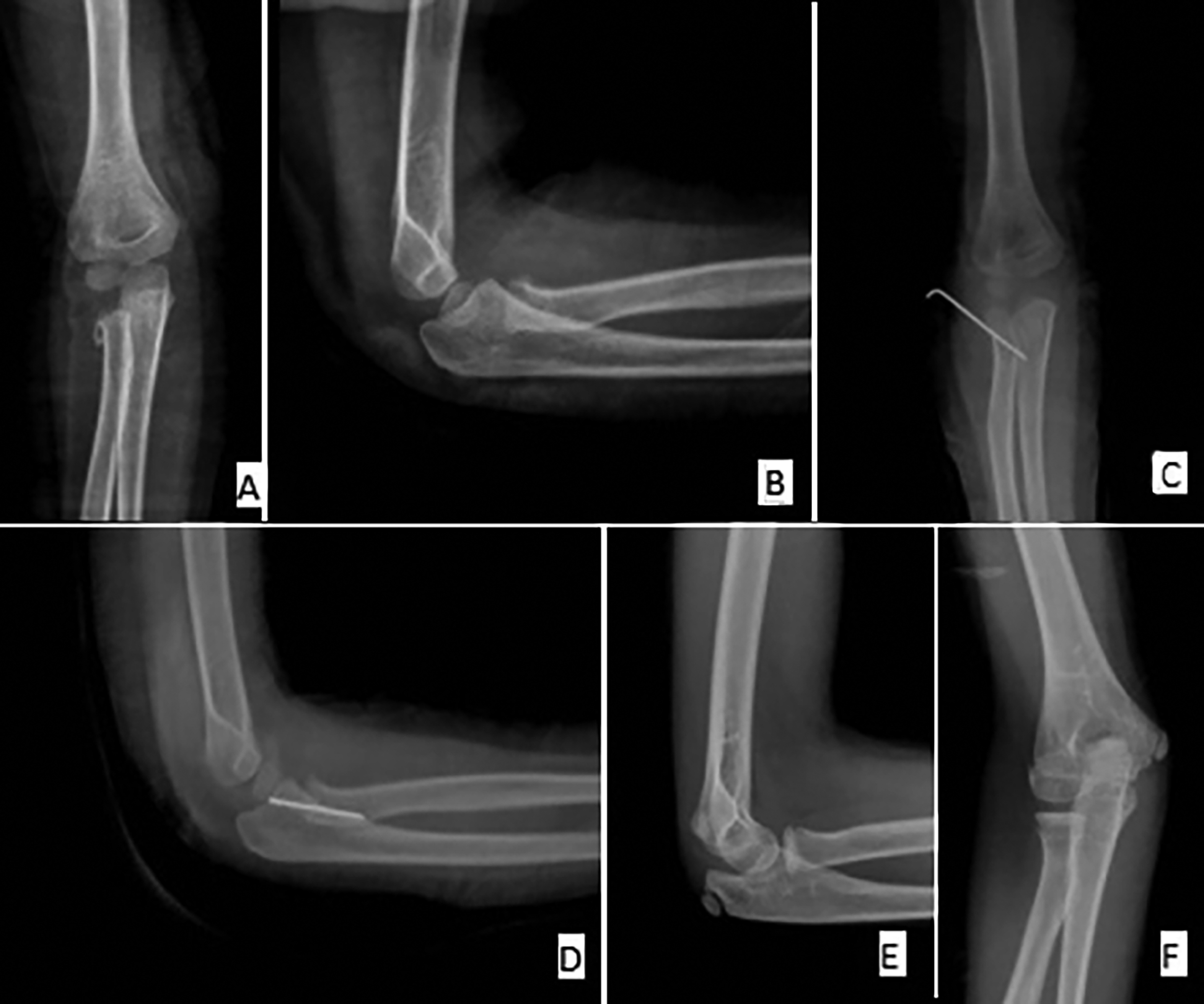 Figure 1: Preoperative, postoperative, and 12-month follow-up radiographs of a patient treated with the open reduction technique. (A) Preoperative anteroposterior roentgenography image showing the displacement of the radial neck. (B) Preoperative lateral roentgenography image showing the displacement of the radial neck. (C) Postoperative anteroposterior roentgenography image showing the open reduction. (D) Postoperative lateral roentgenography image showing the open reduction. (E) Two-year follow-up anteroposterior roentgenography image showing excellent results. (F) Two-year follow-up lateral roentgenography image showing excellent results.
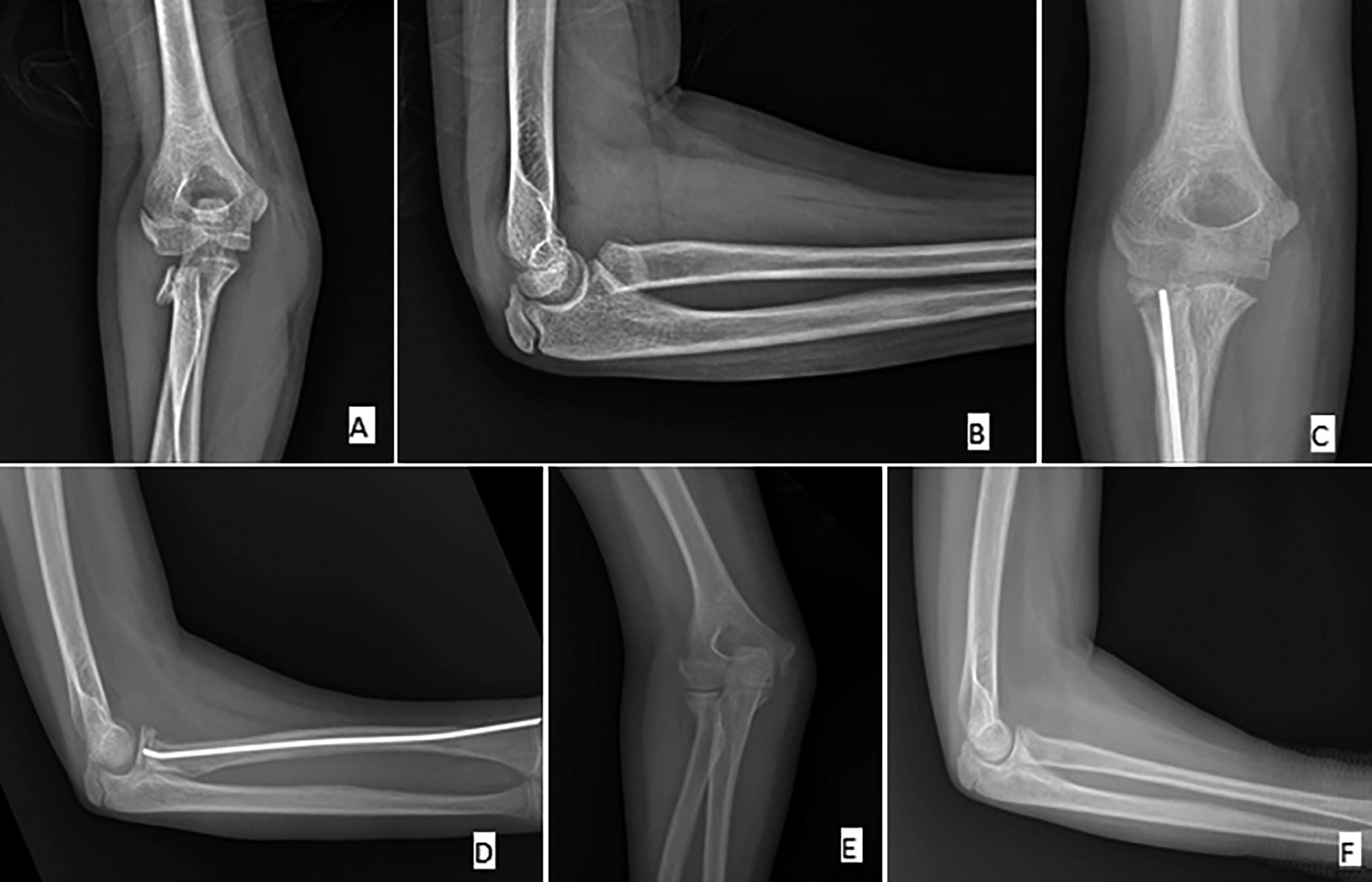 Figure 2. Preoperative, postoperative, and 11-month follow-up radiographs of a patient treated with the Métaizeau technique. (A) Preoperative anteroposterior roentgenography image showing the displacement of the radial neck. (B) Preoperative lateral roentgenography image showing the displacement of the radial neck. (C) Postoperative anteroposterior roentgenography image showing the Métaizeau technique. (D) Postoperative lateral roentgenography image showing the Métaizeau technique. (E) Two-year follow-up anteroposterior roentgenography image showing excellent results. (F) Two-year follow-up lateral roentgenography image showing excellent results.
Posterolateral approach was used after placing the patient placed in the supine position.The anconeus–extensor carpi ulnaris interval was used to expose the orbicular ligament. The joint capsule was cut along the brachioradialis muscle and spatium inter musculare. Fluoroscopy was used to confirm the reduction. A 1.5-mm K-wire used to fix the fracture. The pin was bent and cut, the pintail was retained in the outer skin, and the incision was sutured. After the operation, a long arm cast was applied in all the patients for 4 weeks for pain relief and stabilization. The K-wire was removed after 4 weeks.
Table I: Judet classification.
|
Type |
Angulated angle (°) |
Displacement (%) |
|
I |
No |
No |
|
II |
>30 |
<1/2 of transverse diameter |
|
III |
30-60 |
>1/2 of transverse diameter |
|
IV |
>60 |
Total |
|
Judet IVa: Angulated angle of 60-80°; Judet IVb: Angulated angle >80° |
||
All the patients were followed up regularly for 12 months after surgery. Radiological evaluation was performed using standard anteroposterior and lateral radiographs. Complete fracture healing was defined as full return to activities of daily living and sports. Radiological results were evaluated in accordance with the Ursei Classification.10 MEPS was used to assess functional development.11
Statistical analyses were performed using SPSS software (version 20.0; IBM Corp., Armonk, NY, USA). Descriptive statistics were expressed as means, medians, standard deviations, ranges, and percentages. Data were tested for normality using the Kolmogorov–Smirnov test. A comparative analysis of two independent groups was performed using Pearson’s chi-square test for categorical variables. An independent samples t-test was performed for continuous variables in accordance with normality testing. A two-sided p value of <0.05 was considered significant. By assuming the difference in the large effect size between the groups (effect size = 0.55), the sample size was calculated as 95% power and 43 cases for the alpha significance level of 0.05.
RESULTS
A total of 47 patients were included in this study, including 22 girls and 25 boys. Their mean age at the time of surgery was 8.57 ± 2.3 years (range, 5–14 years). Both groups were comparable in terms of demographic and clinical characteristics (Table II). All the patients were followed up for minimum 12 months (mean, 17.1 ± 3.76 months; range, 12–24 months). Bone union was achieved in all patients within a mean time of 4.2 weeks.
Of the 22 patients treated with closed reduction with percutaneous reduction using K-wire and the Métaizeau technique, one had a superficial wound infection and another had a heterotopic ossification. The superficial wound infection was resolved with a simple course of parenteral antibiotic therapy. Radiological imaging revealed that the closed reduction approach did not affect the clinical results in the patient with heterotopic ossification.
Of the 25 patients treated with open reduction and internal fixation with K-wire, 5 had pin tract infections, 2 had a radial head avascular necrosis and one had a transient posterior interosseous nerve lesion. The pin tract infections were resolved with a simple course of parenteral antibiotic therapy. The neuropraxia was managed nonoperatively and resolved spontaneously within 8 months. The complication rate in the open reduction treatment group was statistically significantly higher than that in the group of patients who received percutaneous reduction using K-wire and the Métaizeau technique (p = 0.014).
The mean MEPS of the patients who received percutaneous reduction using K-wire with the Métaizeau technique was 95.2, with excellent, good, and fair or poor results in 15 (68%), 7 (31%), and none of the patients, respectively. The mean MEPS in the open reduction / K-wire group was 88, with excellent, good, fair, and poor results in 9 (36%), 12 (48%), 4 (16%), and 0 patients, respectively (Table II). The Métaizeau technique group showed statistically significantly better results than the open reduction group in terms of MEPS (p = 0.014).
Postoperative evaluation using the Ursei classification for radiological evaluation, 19 of the 25 patients who underwent the open reduction technique. Nineteen (76%) had excellent results and 6 (24%) had good results. Of the 22 patients who underwent percutaneous reduction using K-wire with the Métaizeau technique, 20 (90.9%) had excellent results and 2 (9.09%) had good results. The Métaizeau technique group showed statistically nonsignificant but proportionally better results than the open reduction group according to the Ursei classification (p = 0.064).
DISCUSSION
The degree of angulation of the fracture determines the treatment plan for PRNFs. Judet Type 1 and 2 fractures can be treated non-operatively with short-term immobilization. The majority of these individuals have a good prognosis and functional outcomes.12,13 The treatment of Judet Type 3 radial neck fractures remains controversial in the literature.14,15 For Judet Type 4 radial neck fractures, surgical treatment is recommended.13 By including only Judet Type 4 fractures in the study, any effect of fracture type on treatment outcomes was eliminated.
A statistically significant difference in MEPS was observed between the two groups in this study. Functional results were better in the group of patients treated with closed reduction and the Métaizeau technique than in the open reduction group. A statistically significant complication rate was observed in the open reduction and K-wire group as compared with the Métaizeau technique group. There was no significant difference between the two groups in terms of neurological complications. The radiological and functional results of the patients in the present study were consistent with previous findings reported in the literature.
Although many studies have focused on the advantages of the closed reduction and Métaizeau techniques,16-21 Sirois et al. reported that undesirable radial head inversion during closed reduction may lead to minor complications.22 In addition, another study reported that closed reduction of Judet type 4 fractures would be difficult; therefore, open reduction would be necessary.
|
Table II: Comparison of the outcomes of two methods for the treatment of Judet Type IV radial neck fractures. |
||||
|
Surgical technique |
Open reduction with K-wire fixation |
Percutaneus ascociated Metaizeau technique |
p-value |
|
|
Age in year Mean ± SD Median (Min_Max) |
8.6 ± 2.3 8 (5-12)
|
8.5 ± 1.5 8 (6-11)
|
0.810 |
|
|
Judet cassification |
Judet type 4A |
18 |
10 |
0.06
|
|
Judet type 4B |
7 |
12 |
||
|
MEPS Mean ± SD Median (Min_Max) |
88.0 ± 10.6 85 (70-100) |
95.2 ± 7.2 100 (85-100) |
,010 |
|
|
Complication |
Pin tract infection |
5 |
1 |
0.014
|
|
PIN deficit |
1 |
0 |
||
|
Avascular necrosis |
2 |
0 |
||
|
Hypertrophic scar |
0 |
0 |
||
|
Heterotopic Ossification |
0 |
1 |
||
|
Operation time mean ± SD |
45.4 ±11.5 |
65.0 ± 10.0 |
<0.001 |
|
|
PIN: Posterior interosseous nerve; MEPS: Mayo elbow performance score. |
||||
In this study, radial head inversion did not occur as a complication in the closed reduction group. In addition, there was no case of failed closed reduction. therefore, open reduction was needed. Hence, we argue that closed reduction should be performed as the initial treatment for Judet type 4 fractures, and open reduction should be started only if closed reduction is unsuccessful.
Along with minimally invasive surgical treatments being in the forefront of orthopedic surgery, the use of fluoroscopy has been increasing. In this study, the operation time was significantly higher in the closed reduction method than in the open reduction method. Owing to the difficulty of achieving closed reduction, radiation exposure was increased. Lee et al. argued that ultrasonography (USG)-guided reduction is more reliable.23 In addition, studies have described arthroscopy-assisted reduction in recent years.8,24 Although USG- and arthroscopy-assisted reduction are advantageous in terms of reduced radiation exposure, their use in daily clinical practice is not yet practical, and closed reduction is still the most effective and safest method in terms of reduced risk of complications.
This study has some strengths and limitations. The strongest aspect of this study is the comparison of two different methods in the surgical treatment of radial neck fractures with similar patient distributions in the two groups. The disadvantages are a limited sample size and retrospective study design.
CONCLUSION
Closed reduction using the Métaizeau technique with the elastic stable intramedullary nailing method satisfies all the criteria for minimally invasive bone surgery. Regarding treatment of PRNF in children, this approach has been shown to be considerably efficient, with excellent functional and esthetic outcomes and a low rate of complications if the indications and biomechanical principles are considered.
ETHICAL APPROVAL:
This study was conducted retrospectively in accordance with the ethical standards of the SBU Haseki Training and Research Hospital Clinical Research Ethics Committee and the 1975 Declaration of Helsinki revised in 2013. Ethics committee approval was obtained (Decision No. 2020-27, 25/11/2020).
PATIENTS' CONSENT:
Consent was obtained from the parents of the patients as "I voluntarily accept the participation of my child in the clinical trial".
COMPETING INTEREST:
None of the authors received any type of financial support that could be considered a potential conflict of interest regarding the manuscript or its submission.
AUTHORS’ CONTRIBUTION:
MY: Conception and design, drafting the article.
MA: Data acquisition, analysis and interpretation of data.
MM: Drafting the article; revising it critically for important intellectual content in the article.
All authors approved the final version of the manuscript to be published.
REFERENCES
- Landin LA, Danielsson LG. Elbow fractures in children. An epidemiological analysis of 589 cases. Acta Orthop Scand 1986; 57(4):309-12. doi:10.3109/17453678608994398.
- Chambers HG. Fractures of the proximal radius and ulna. In: Beaty JH, Kasser JR, eds. Fractures in Children. Philadelphia, PA: Lippincott Williams and Wilkins; 2001:483–528.
- D'souza S, Vaishya R, Klenerman L. Management of radial neck fractures in children: A retrospective analysis of one hundred patients. J Pediatr Orthop 1993; 13(2):232-8.
- Neher CG, Torch MA. New reduction technique for severely displaced pediatric radial neck fractures. J Pediatr Orthop 2003; 23(5):626-8. doi:10.1097/00004694-200309000- 00009.
- Vahvanen V, Gripenberg L. Fracture of the radial neck in children. A long-term follow-up study of 43 cases. Acta Orthop Scand 1978; 49(1):32-8. doi:10.3109/1745367780900 5720.
- Schmittenbecher PP, Haevernick B, Herold A, Knorr P, Schmid E. Treatment decision, method of osteosynthesis, and outcome in radial neck fractures in children: A multicenter study. J Pediatr Orthop 2005; 25(1):45-50. doi:10. 1097/00004694-200501000-00011.
- Du X, Yu L, Xiong Z, Chen G, Zou J, Wu X, et al. Percutaneous leverage reduction with two Kirschner wires combined with the Métaizeau technique versus open reduction plus internal fixation with a single Kirschner-wire for treating Judet IV radial neck fractures in children. J Int Med Res 2019; 47(11):5497-507. doi:10.1177/03000605198 25990.
- Colozza A, Padovani S, Caruso G, Cavaciocchi M, Massari L. Arthroscopically-assisted reduction and pinning of a radial neck fracture in a child: A case report and review of the literature. J Med Case Rep 2020; 14(1):78. doi:10.1186/ s13256-020-02390-0.
- Judet J, Judet R, Lefranc J. Fracture of the radial head in the child. In: Ann Chir 1962; 16:1377-85.
- Pring ME. Pediatric radial neck fractures: When and how to fix. J Pediatr Orthop 2012; 32 Suppl 1:S14-S21. doi:10. 1097/BPO.0b013e31824b251d.
- Ursei M, Sales de Gauzy J, Knorr J, Abid A, Darodes P, Cahuzac JP. Surgical treatment of radial neck fractures in children by intramedullary pinning. Acta Orthop Belg 2006; 72(2):131-7.
- Morrey BF, An KN, Chao EYS. Functional evaluation of the elbow. In: Morrey BF, ed. The Elbow and Its Disorders. 2nd ed. Philadelphia: WB Saunders; 1993: 86e89.
- Fowles JV, Kassab MT. Observations concerning radial neckfractures in children. J Pediatr Orthop 1986; 6:51-7.
- Yarar S, Sommerfeldt DW, Gehrmann S, Rueger JM. Stark dislozierte radiushalsfrakturen nach minimal-invasiver joystick-reposition und prévot-nagelung: Langzeitverlauf im kindesalter [severely displaced radial neck fractures after minimally invasive joystick reduction and prévot nailing: Long-term course in childhood]. Unfallchirurg 2007; 110(5):460-6. doi:10.1007/s00113-006-1181-7.
- Eberl R, Singer G, Fruhmann J, Saxena A, Hoellwarth ME. Intramedullary nailing for the treatment of dislocated pediatric radial neck fractures. Eur J Pediatr Surg 2010; 20(4): 250-2. doi:10.1055/s-0030-1249104.
- Kruppa C, Königshausen M, Schildhauer TA, Dudda M. Isolated pediatric radial head and neck fractures. A rare injury. Analysis and follow up of 19 patients. Injury 2015; 46 Suppl 4:S10-S6. doi:10.1016/S0020-1383(15)30013-9.
- Bither N, Gupta P, Jindal N. Pediatric displaced radial neck fractures: REtrospective results of a modified Metaizeau technique. Eur J Orthop Surg Traumatol 2015; 25(1): 99-103. doi:10.1007/s00590-014-1452-x.
- Tarallo L, Mugnai R, Fiacchi F, Capra F, Catani F. Management of displaced radial neck fractures in children: Percutaneous pinning vs. elastic stable intramedullary nailing. J Orthop Traumatol 2013; 14(4):291-7. doi:10.1007/s10195- 013-0252-0.
- Trabelsi A, Khalifa MA, Brahem R, Jedidi M, Bouattour K, Osman W, et al. Radial neck fracture in children: Anatomic and functional results of Metaizeau technique. Pan Afr Med J 2020; 36:144. doi:10.11604/pamj.2020.36.144.22971.
- Bilal O, Murat Kalender A, Karsli B, Kilinçoğlu V, Kinaş M, Dundar N. Radiological and functional outcomes of modified Metaizeau technique in displaced radial neck fractures. Acta Orthop Belg 2021; 87(2):235-241.
- Çevik N, Cansabuncu G, Akalın Y, Otuzbir A, Oztürk A, Ozkan Y. Functional and radiological results of percutaneous K-wire aided Métaizeau technique in the treatment of displaced radial neck fractures in children. Acta Orthop Traumatol Turc 2018; 52(6):428-434. doi:10.1016/j.aott.2018.07.007.
- Sirois ZJ, Kreul SM, Shank CF. Inadvertent radial head inversion during closed reduction of a pediatric radial neck fracture. J Am Acad Orthop Surg 2019; 27(9):e414-e7. doi:10. 5435/JAAOS-D-17-00668.
- Lee JE, Kim JB, Choi ES. Ultrasonography-guided reduction of pediatric radial neck fractures. BMC Musculoskelet Disord 2017; 18(1):516. doi:10.1186/s12891-017-1891-8.
- Kim JY, Kim JW, Lee JM, Lee SH, Cho HH, Han JK, et al. Arthroscopic treatment of radial neck fractures in children: A technical note. Arthroscopy and Orthopedic Sports Medicine 2017; 4(1):39-43.