Fibrolipoma of Tongue: A Rare Entity
By Salima Mansoor1, Nausheen Yaqoob1, Anila Haneef1, Sunder Sham1, Asif Ali Arain2Affiliations
doi: 10.29271/jcpsp.2022.06.814ABSTRACT
Lipomas are benign mesenchymal tumors that comprise almost one-half of all soft tissue tumors. Lipomas can occur at any site where fat cells are present but are rare in the oral cavity, especially the tongue, which is a very rare site. Lipoma has several variants. Fibrolipoma is a rare variant and accounts for 25–40% of lipomas of the tongue. In only 14 cases, the diagnosis of fibrolipoma has been made histologically. Most cases of lipomas occur above 40 years of age. Herein, we report a case of fibrolipoma of the tongue in a young female of 18 years. The patient presented with complaint of swelling on the dorsum of the tongue for 8 years. The swelling was surgically excised and microscopically a diagnosis of fibrolipoma was made. In conclusion, oral lipomas especially lingual lipomas are a rare entity. Although oral lipomas mostly occur above 40 years of age, they can occur at a younger age.
Key Words: Fibrolipoma, Tongue, Histopathology.
INTRODUCTION
Lipomas are benign mesenchymal tumors that are very common and are comprised of mature adipocytes. They usually grow slowly and form a lobulated mass surrounded by a capsule. They can arise from almost all organs in the body where fat cells are present and may be well encapsulated or appear as infiltrating lesions.1,2 Lipomas rarely occur in the oral cavity. The tongue is an infrequent site for lipomas.2 Of all tongue tumors, lipomas account for only 0.3% of cases.3 Lipomas of the tongue may develop as a solitary lesion or can occur as a manifestation of syndromes such as Gardner’s syndrome or hereditary multiple lipomatosis.1,4 There are several variants of lipoma.2 In contrast to simple/conventional lipoma, other variants of lipoma occur less frequently.5 Fibrolipoma is also a rare variant that sometimes may cause suspicion of infiltrating lesions.5 It can involve various sites in the oral cavity including lips, buccal mucosa, palate, and tongue.6,7 In this case report, we present a case of fibrolipoma of the tongue in a young girl where the diagnosis was solely made through histopathological examination.
CASE REPORT
An 18-year female was referred from family medicine to Otolaryngology department with the complaint of swelling on the dorsum of the tongue for 8 years. The swelling was painless, pink in color, non-tender and gradually increased in size.
The patient did not have a significant past medical or surgical history. Family history was also unremarkable for any tumors. The patient did not seek any treatment in the past as the swelling was painless and the patient’s family had a low socioeconomic status.
On examination, a well-demarcated swelling was identified which was pink in color, measuring approximately 3 × 3 cm with no ulceration or inflammation (Figure 1). The swelling was non-tender and firm on palpation. There was no palpable lymphadenopathy. The differentials included hemangioma, lymphangioma, and mucocele. A decision was made to excise the lesion.
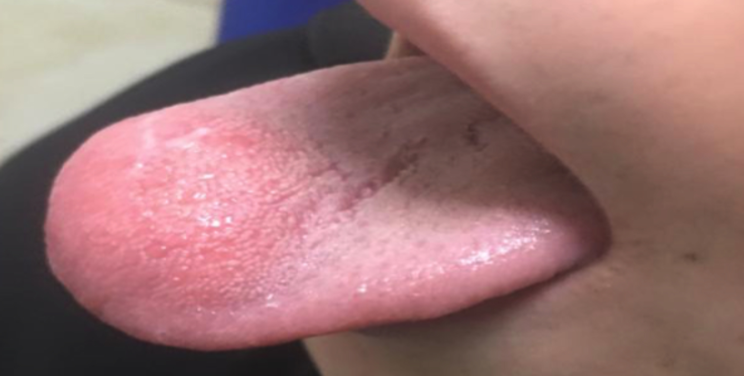 Figure 1: Mass located at the anterior third of the tongue.
Preoperative investigations including complete blood count, serum electrolytes, PT, APTT, renal function tests, and blood glucose levels were all within normal limits. The patient was operated and the lesion was shelled out with no adhesion to the surrounding structures. Post-operatively, the patient remained stable and did not develop any postoperative complications.
Gross examination of the lesion showed a well-circumscribed mass measuring 3.5 × 2.5 × 1.5 cm. On sectioning, the cut surface was firm, grey-white with no areas of hemorrhage or necrosis.
Microscopy revealed an adipocytic lesion comprising of lobules of adipocytes separated by thick intervening fibrous bands (Figure 2). No lipoblasts were identified. The salivary gland was identified at the periphery. The features were consistent with fibrolipoma of the tongue.
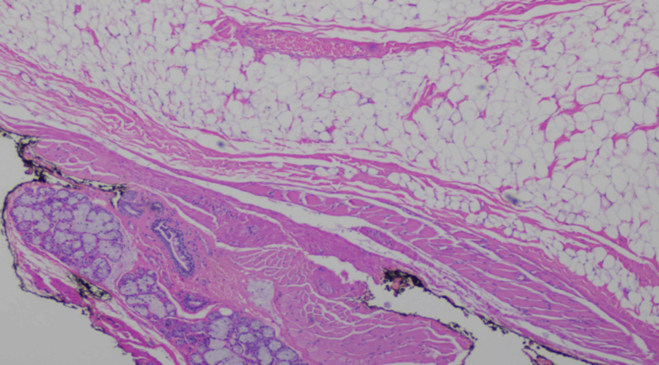 Figure 2: Section showing fibrolipoma with skeletal muscle fibers of tongue and minor salivary gland tissue at the periphery (H&E, ×20).
DISCUSSION
Lipomas can involve almost all sites that have adipose tissue but are rare in the oral cavity and maxillofacial areas.2,6 A study by Fregnani et al. reviewed 46 cases of oral cavity lipomas between 1970-2001, which represented 0.5% of all tumors seen in their Oral pathology department.2 Oral lipomas that are superficial may appear yellow and therefore are easy to diagnose but deep-seated oral lipomas appear pink8 and seldom diagnosed early as they are often asymptomatic till they attain a considerable size.1,2,6 In such situations, they may be misdiagnosed clinically as hemangiomas. Other differentials include mucocele, lymphangioma, neuroma, rhabdomyoma, neurofibroma, or salivary gland tumors.2 Tongue lipomas are seen in all ages but are more frequent in adults >40 years.1,2,6 A case report and literature review by Lu et al. reported that tongue lipomas were seen in patients between the ages of 1-66 years. Out of 18 cases reported, only two cases were seen below 1 year of age and 16 cases were seen in patients above 30 years of age. However, no cases were reported between the ages of 1 and 30 years.1 Another study reported that lipomas most commonly occur in patients older than 60 years and occurrence in children is rare. In their study, the mean age of patients with oral lipomas was 59 years and only one case occurred in <40 years.9 Fibrolipoma is a rare variant and accounts for 25–40% of lipomas of the tongue.5,10 In only 14 cases, the diagnosis of fibrolipoma has been made histologically.5
The etiology and pathogenesis of lipomas are unknown but various theories include repeated trauma, infections, altered lipid metabolism, hormonal problems, degeneration of fat, and ectopic fetal fat tissue.1,9 Preoperatively, the diagnosis can be assisted by ultrasound, computed tomography (CT) scan, or magnetic resonance imaging (MRI).2 Sometimes aspiration of the lesion with a needle can be performed to differentiate between hemangioma, mucocele, or lymphangioma. Definitive diagnosis requires Fine needle aspiration (FNAC), incisional or excisional biopsy followed by histopathological examination.2 Treatment of all types of lipoma is surgical excision regardless of the histological type. Recurrence is rarely reported.2 Excision of the capsule of lipoma is necessary to prevent a recurrence.
In conclusion, oral lipomas, especially lingual lipomas, are a rare entity. Although oral lipomas mostly occur above 40 years of age, they can occur at a younger age.
PATIENT’S CONSENT:
Written informed consent was obtained from a legally authorized representative(s) for anonymized patient information to be published in this article.
COMPETING INTEREST:
The authors declared no potential conflicts of interest with respect to the research, authorship and/or publication of this article.
AUTHORS’ CONTRIBUTION:
NY, AH, SS: Performed the histological examination, of specimen.
SM, SS, AH: Contributed in writing the manuscript.
AAA:
All authors approved the final version of the manuscript to be published.
REFERENCES
- Lu SL, Zhen JJ, Wu H, Li T, Dong G, Wang YL, et al. Tongue lipoma in an older male: A case report and literature review of patients with tongue lipoma reported in China. Oncol Lett 2016; 11(1):419-22. doi: 10.3892/ol.2015.3865.
- Magadum D, Sanadi A, Agrawal JM, Agrawal MS. Classic tongue lipoma: A common tumour at a rare site. BMJ Case Rep 2013; 2013:p.bcr2012007987. doi: 10.1136/bcr- 2012-007987.
- Srinivasan K, Hariharan N, Parthiban P, Shyamala R. Lipoma of tongue - A rare site for a rare site for a common tumour. Indian J Otolaryngol Head Neck Surg 2007; 59(1):83-4. doi: 10.1007/s12070-007-0027-0.
- Mungul S, Maharaj S, Masege SD. Lingual Fibrolipoma - A rare clinicopathological entity. S Afr J Surg 2017; 55:36-8.
- Laconetta G, Friscia M, Cecere A, Romano A, Orabona GD, Califano L. Rare fibrolipoma of the tongue: A case report. J Med Case Rep 2015; 9(1):177. doi: 10.1186/s13256- 015-0653-1.
- Pereira T, Shetty S, Sapdhare S, Tamgadge A. Oral fibrolipoma: A rare histological variant. Indian J Dent Res 2014; 25(5):p.672-4. doi: 10.4103/0970-9290.147123.
- Gujjari SK, Shah M, Hedge U, Doddawad VG. Fibrolipoma: Report of two intraoral cases. J Clin Diagn Res 2012; 6(3): 524-6. doi: JCDR/2012/3504:1946.
- Wu YH, Lin PY, Chang MH, Chiang CP. Lipoma of the tongue. J Formos Med Assoc 2017; 116(12):1006-7. doi: 10.1016/j.jfma.2017.11.002.
- Naruse T, Yanamoto S, Yamada S, Rokutanda S, Kawakita A, Takahashi H, et al. Lipomas of the oral cavity: Clinicopathological and immunohistochemical study of 24 cases and review of the literature. Indian J Otolaryngol Head Neck Surg 2015; 67(Suppl 1):67-73. doi: 10.1007/ s12070-014-0765-8.
- Baonerkar HA, Vora M, Sorathia R, Shinde S. The lipoma of tongue - A rare site for a tumor: Case report and review of the literature. Indian J Dent 2015; 6(4):207-10. doi: 10.4103/0975-962X.168520.