Different Translocations of Multiple Myeloma on Fluorescent In Situ Hybridization (FISH) with Clinical Correlation
By Muhammad Umar, Hamid Saeed Malik, Baber Zaman, Muhammad Waleed Ahmad, Fauzia Khan, Hira NadeemAffiliations
doi: 10.29271/jcpsp.2023.03.281ABSTRACT
Objective: To evaluate the association of chromosomal translocations in multiple myeloma (MM) detected by Fluorescent In Situ Hybridization (FISH) and its clinical characteristics.
Study Design: Cross-sectional study.
Place and Duration of the Study: Department of Haematology, Armed Forces Institute of Pathology (AFIP), Rawalpindi, Pakistan, from February to August 2022.
Methodology: A total of 40 cases of MM were included. All cases were diagnosed using international myeloma working group (IMWG) criteria. Clinical presentations like bone pain, backache, fatigue, pallor, and weight loss were noted. The workup for myeloma-defining events was done. FISH analysis was done for t (4;14), t (11,14), t (14;16), t (14;20), and del 17p. Data were analysed using the chi-square test. A p-value ≤0.05 was considered statistically significant.
Results: Out of 40 patients, 8 (20%) were females and 32 (80%) were males. The highest frequency of cases were noted among males in the age group >60 years and females in the age group 40-60 years. FISH for t (4;14) was positive in 22 (55%) patients, for t (11;14) was positive in 4 (10%) patients, for t (14;16) was positive in 3 (7.5%) patients, and for t (14;20) was positive in 3 (7.5%) patients, while for del17p was positive in 8 (20%) patients. Cases with t (4;14), t (11;14), and t (14;20) had bone pain, fatigue, and backache as the most common presentations. Among the various parameters studied, lytic lesions, beta-2 microglobulin, spike protein, deranged haemoglobin, TLC, ESR, albumin, and creatinine were significant risk factors in patients who were tested positive for various mutations.
Conclusion: The FISH technique has brought an immense uprising in the genetic analysis of MM. Among translocations, t (4;14) and del17p are associated with poor clinical outcomes and prognosis. If the diagnosis of MM is delayed, then an increase in morbidity and mortality can occur.
Key Words: Multiple myeloma, FISH, Translocations.
INTRODUCTION
An abnormal proliferation of monoclonal immunoglobulin-producing plasma cells in the bone marrow causes multiple myeloma (MM). This haematological malignancy can result in anaemia, hypercalcemia, osteolysis, osteopenia, and fractures due to uncontrolled plasma cell proliferation.1 MM is a comparatively uncommon cancer and accounts for approximately 1 to 2% of all cancers, and 17% of haematological malignancies. It is more prevalent among males than females.
Among different populations of the world, it is relatively higher among African American descent.2 Annual death attributed to MM worldwide is approximately 117,000 out of 180,000 cases.3 Patients with monoclonal gammopathy of undetermined significance (MGUS) are at more risk of MM if exposed to agent orange.4 Another risk factor is family history. The risk of developing MM is 3.7-fold higher for persons with a first degree relative to MM.5
Cases with MM present with symptoms related to the infiltration of plasma cells into bone or kidney, or other organs. Myeloma-defining events include hypercalcemia, renal insufficiency, anaemia, and bone disease. Non-specific symptoms include weight loss, backache, and fatigue.6 Other benign causes like monoclonal gammopathy of undetermined significance (MGUS), Walderstrom macroglobulinemia, solitary plasmacytoma, amyloidosis, and light chain/ heavy chain deposition disease should be differentiated from MM.7
The genetic event concerning the progression of the disease is important in understanding disease pathogenesis and may then be used to predict new treatment strategies.8 Genome-wide association study (GWAS) suggested that persons with a common variation of 3p22.1 or 7p15.3 loci are at a higher risk of developing MM and with clinically severe disease.9 MM is genomically very unstable, and it is characterised by translocations involving the IGH locus located on chromosome 14q32, methylation, hypoploidy or hyperploid, and dysregulation of cyclin D genes. Half of the cases show IGH translocations, which include recurrent five chromosomal patterns t (4;11), t (4;14), t (14;16), t (6;14) and t (14;20).10 In Pakistan, data on these translocations is very sparse. So, this study was conducted to determine different translocations of MM using Fluorescent In Situ Hybridization (FISH) and correlate it with the clinical presentation.
METHODOLOGY
This cross-sectional study was conducted at the Department of Haematology, Armed Forces Institute of Pathology (AFIP), Rawalpindi, Pakistan, from February to August 2022. Ethical approval was obtained from the institutional review board (IRB) vide reference number (FC-HEM21-13/READ-IRB/22/1292). After a thorough literature search, a sample size of 36 was calculated using a WHO calculator, keeping a 5% margin of error, 95% confidence level, and a prevalence of 2.4%.11 Sampling was done using the non-probability convenience sampling technique. All available cases n=40 during the study period were recruited.
Newly diagnosed cases of multiple myeloma (diagnosed according to the IMWG diagnostic criteria) were included in the study.12 Exclusion criteria were patients with diagnosed MGUS and other plasma disorders. Patients already on treatment were also excluded from the study. Before enrolling all patients, their written consent was obtained, and the confidentiality of the patients was ensured at all levels.
Detailed history and complete physical examination were done. Complete blood counts were performed on the Sysmex XN-3000. Baseline investigations were carried out. Serum and urine protein electrophoresis and immunofixation were also done. Each patient was tested for RFTS, serum calcium, serum albumin, and beta-2 microglobulins. Bone marrow aspiration and trephine biopsy were done to see plasma cell numbers and morphology. The diagnosis of multiple myeloma was made as per the IMWG diagnostic criteria of MM.
Blood or bone marrow samples were analysed for interphase FISH studies. Probes used for cytogenetic translocation on FISH were t (4;14), t (6;14), t (11;14), t (14;16), t (14;18), t (14;20), del17p, 1p32, and 1q21, which affect the FGFR3 gene, upregulates cyclin D1, deregulate c-MAF and MAFB, and inactivate p53 tumour, respectively. Suppressor gene samples were processed by standard methods for culture.
A 3 ml of sample was collected in sodium heparin and 0.5 ml of blood is added to 7 ml of culture media and incubated at 37OC for 24 hours. After 24 hours, 2.5 ml Colchicine was added and incubated at 37OC for 45 min, then centrifuged, and supernatant was removed. Distilled water and KCL were added and incubated for 10 min, recentrifuged, and supernatant were discarded. Sample was fixed with glacial acetic acid. Repeated washings were done. The slide was prepared with a pellet, fixed with 20 SSC for 2 min and then with ethanol. The slide was dried and FISH probe was applied. Denaturation at 74OC for 15 min, hybridisation at 37OC for 18 hours, then washed with alcohol followed by 10% SSC solution, counterstained with Dappiz stain and placed at 20OC for 30 min. In each probe set, 500 nuclei were analysed under spectrum filter using a fluorescence microscope. t (4;14), t (6;14), t (11;14), t (14;16), t (14;18), t (14;20), 17p/13, 1p32, and 1q21 were used as fusion probes. The FISH analysis is shown in Figure 1.
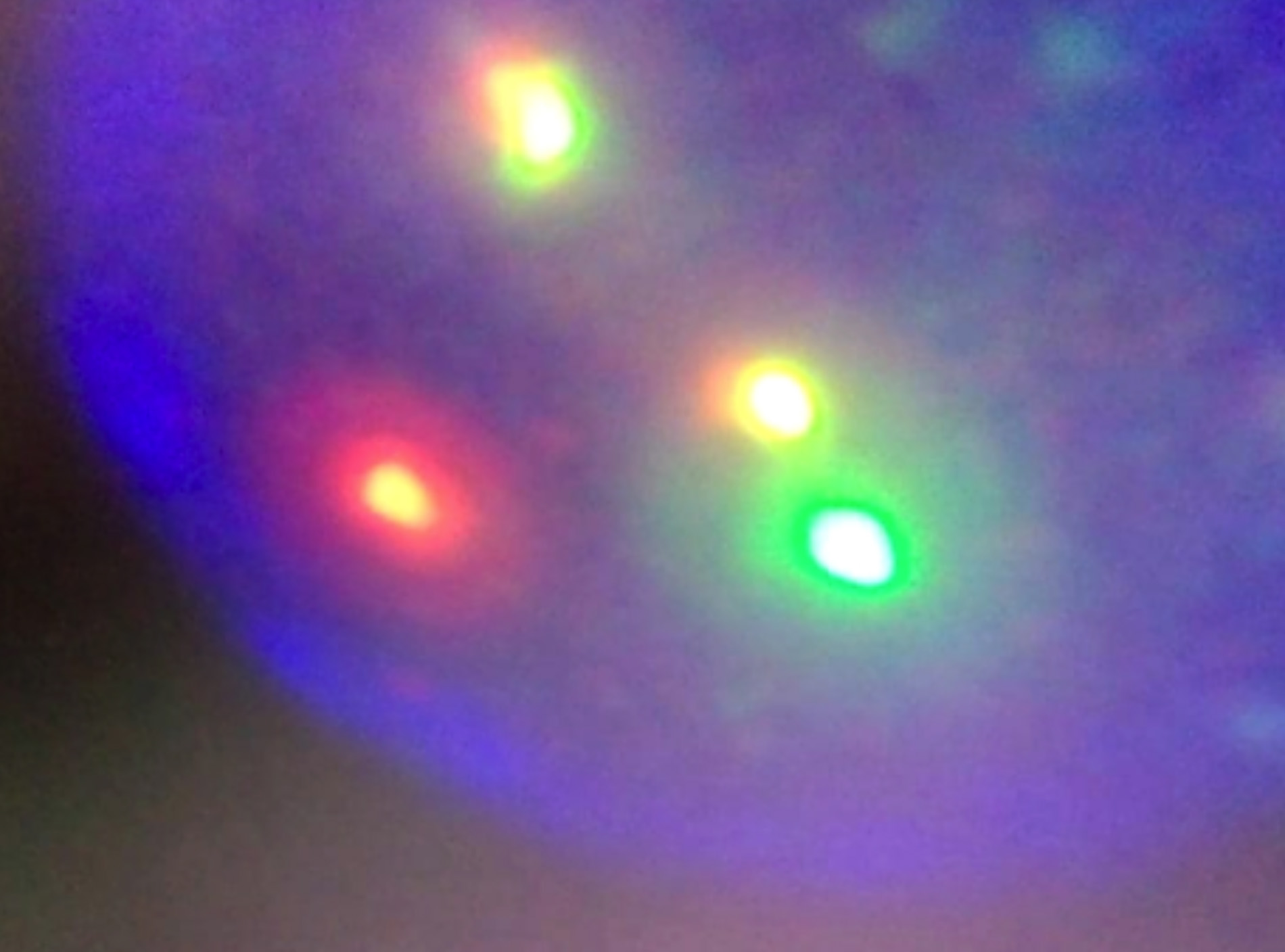 Figure 1: FISH analysis. (The orange labelled probes indicate a breakpoint at the FGFR3 gene and the green probes indicate a breakpoint at 14q32 proximal and distal to the IGH gene region. One green, one orange, and two green-orange fusion signals indicate t (4;14)).
Data were entered in Microsoft excel and later analysed using statistical package for social sciences (SPSS) 21.0. Descriptive statistics were expressed as mean ± standard deviation (SD) and categorical data were presented as frequency and percentage. A chi-square test was applied. A p-value ≤0.05 was considered statistically significant.
RESULTS
Out of the 40 patients, 8 (20%) were females and 32 (80%) were males. The mean age of patients was 61.00 ±9.716 years. The highest frequency of cases were noted among males in the age group >60 years and among females in the age group 40-60 years.
FISH for t (4;14) was positive in 22 (55%) patients, for t (11;14) was positive in 4 (10%) patients, for t (14;16) was positive in 3 (7.5%) patients and for t (14;20) was positive in 3 (7.5%) patients, while for del17p was positive in 8 (20%) patients.
Cases with t(4;14), t(11;14), and t(14;20) had bone pain, fatigue, and backache as the most common presentations. However, cases with del17p were presented frequently with bone pain, fatigue, backache, and weight loss. Different signs, symptoms, and chromosomal translocations were noted on FISH are shown in Table I.
Table I: Signs, symptoms, and chromosomal translocations noted on FISH using chi-square test.|
Signs and Symptoms |
Chromosomal translocations detected on FISH |
p-value |
||||
|
t(4;14) |
t(11;14) |
t(14;16) |
t(14;20) |
del17p |
||
|
Bone pain, fatigue, and backache |
13 (59.1%) |
3 (75.0%) |
1 (33.3%) |
2 (66.7%) |
2 (25.0%) |
0.461 |
|
Bone pain, fatigue, and weight loss |
1 (4.5%) |
0 (0.0%) |
0 (0.0%) |
0 (0.0%) |
0 (0.0%) |
|
|
Bone pain, fatigue, backache, and weight loss |
3 (13.6%) |
0 (0.0%) |
0 (0.0%) |
0 (0.0%) |
3 (37.5%) |
|
|
Fatigue, backache, and pallor |
2 (9.1%) |
0 (0.0%) |
1 (33.3%) |
0 (0.0%) |
0 (0.0%) |
|
|
Bone pain, fatigue, backache, and pallor |
1 (4.5%) |
0 (0.0%) |
0 (0.0%) |
0 (0.0%) |
0 (0.0%) |
|
|
Bone pain, fatigue, backache, pallor, and weight loss |
1 (4.5%) |
1 (25.0%) |
0 (0.0%) |
0 (0.0%) |
1 (12.5%) |
|
|
Backache, pallor, and weight loss |
1 (4.5%) |
0 (0.0%) |
1 (33.3%) |
0 (0.0%) |
0 (0.0%) |
|
|
Bone pain, backache, and weight loss |
0 (0.0%) |
0 (0.0%) |
0 (0.0%) |
1 (33.3%) |
2 (25.0%) |
|
|
*p-value was calculated by using the chi-square test. |
||||||
Table II: Chromosomal translocations and laboratory parameters studied using chi-square test.
|
|
|
Chromosomal translocations detected on FISH |
p-value |
||||
|
|
t (4;14) |
t (11;14) |
t (14;16) |
t (14;20) |
del17p |
||
|
Family history of haematological malignancy |
Yes |
0 (0.0%) |
0 (0.0%) |
0 (0.0%) |
1 (33.3%) |
0 (0.0%) |
0.013 |
|
No |
22 (100.0%) |
4 (100.0%) |
3 (100.0%) |
2 (66.7%) |
8 (100.0%) |
||
|
Platelets count |
<150 x 109/L |
5 (22.7%) |
1 (25.0%) |
1 (33.3%) |
2 (66.7%) |
4 (55.0%) |
0.437 |
|
>150 x 109/L |
17 (77.3%) |
3 (75.0%) |
2 (66.7%) |
1 (33.3%) |
4 (55.0%) |
||
|
Lytic lesion on skeleton survey |
Solitary lesion |
1 (4.5%) |
0 (0.0%) |
3 (100.0%) |
0 (0.0%) |
0 (0.0%) |
<0.001 |
|
Multiple lytic lesion |
19 (86.4%) |
3 (100.0%) |
0 (0.0%) |
1 (33.3%) |
2 |
||
|
Compression Fracture |
2 (9.1%) |
0 (0.0%) |
0 (0.0%) |
2 (66.7%) |
6 |
||
|
Serum beta-2 microglobulin |
<3.5 ug/mL |
0 (0.0%) |
0 (0.0%) |
1 (33.3%) |
0 (0.0%) |
0 (0.0%) |
<0.001 |
|
3.5-5.5 ug/mL |
0 (0.0%) |
2 (50.0%) |
2 (66.7%) |
0 (0.0%) |
1 (12.5%) |
||
|
>5.5 ug/mL |
22 (100.0%) |
2 (50.0%) |
0 (0.0%) |
3 |
7 (87.5%) |
||
|
Serum albumin |
<3.5 g/dL |
0 (0.0%) |
2 (50.0%) |
2 (66.7%) |
0 (0.0%) |
1 (12.5%) |
0.002 |
|
>3.5 g/dL |
22 (100.0%) |
2 (50.0%) |
1 (33.3%) |
3 (100.0%) |
7 (87.5%) |
||
|
ISS stage |
I |
0 (0.0%) |
0 (0.0%) |
1 (33.3%) |
0 (0.0%) |
0 (0.0%) |
0.001 |
|
II |
0 (0.0%) |
1 (25.0%) |
2 (66.7%) |
0 (0.0%) |
1 (12.5%) |
||
|
III |
20 (100.0%) |
3 (75 .0%) |
0 (0.0%) |
3 (100.0%) |
7 (87.5%) |
||
|
Serum M spike |
<1 g/dL |
0 (0.0%) |
0 (0.0%) |
1 (33.3%) |
0 (0.0%) |
0 (0.0%) |
0.013 |
|
>1 g/dL |
22 (100.0%) |
4 (100.0%) |
2 (66.7%) |
3 (100.0%) |
8 (100.0%) |
||
|
Immunoelectrophoretic abnormality |
IgG |
22 (100.0%) |
3 (75.0%) |
1 (33.3%) |
2 (66.7%) |
5 (62.5%) |
0.007 |
|
IgA |
0 (0.0%) |
1 (25.0%) |
2 (66.7%) |
0 (0.0%) |
2 (25.0%) |
||
|
Free light chains |
0 (0.0%) |
0 (0.0%) |
0 (0.0%) |
1 (33.3%) |
1 (12.5%) |
||
|
Serum calcium |
<11 mg/dL |
17 (77.3%) |
3 (75.0%) |
3 (100.0%) |
0 (0.0%) |
5 (62.5%) |
0.059 |
|
>11 mg/dL |
5 (22.7%) |
1 (25.0%) |
0 (0.0%) |
3 (100.0%) |
3 (37.5%) |
||
|
Total leukocyte count |
<11 x 109/L |
2 (9.1%) |
0 (0.0%) |
0 (0.0%) |
0 (0.0%) |
6 (75.0%) |
0.001 |
|
>11 x 109/L |
20 (90.9%) |
4 (100.0%) |
3 (100.0%) |
3 (100.0%) |
2 (25.0%) |
||
|
Haemoglobin |
<10 g/dL |
22 (100.0%) |
4 (100.0%) |
2 |
3 (100.0%) |
8 (100.0%) |
0.013 |
|
>10 g/dL |
0 (0.0%) |
0 (0.0%) |
1 (33.3%) |
0 (0.0%) |
0 (0.0%) |
||
|
Percentage of plasma cells on bone marrow biopsy |
15-30 |
9 (40.9%) |
3 (75.0%) |
3 (100.0%) |
1 (33.3%) |
6 (75.0%) |
0.146 |
|
31-45 |
13 (59.1%) |
1 (25.0%) |
0 (0.0%) |
2 (66.7%) |
1 (12.5%) |
||
|
46-60 |
0 (0.0%) |
0 (0.0%) |
0 (0.0%) |
0 (0.0%) |
1 (12.5%) |
||
|
Erythrocyte sedimentation rate (mm/hr) |
51-70 |
0(0.0%) |
0 (0.0%) |
2 (66.7%) |
0 (0.0%) |
0 (0.0%) |
0.001 |
|
71-90 |
13 (59.1%) |
3 (75.0%) |
1 (33.3%) |
2 (66.7%) |
4 (50.0%) |
||
|
91-110 |
9 (40.9%) |
1 (25.0%) |
0 (0.0%) |
1 (33.3%) |
4 (50.0%) |
||
|
Serum creatinine (µmol/L) |
30-75 |
21 (95.5%) |
4 (100.0%) |
1 (33.3%) |
2 (66.7%) |
6 (75.0%) |
<0.001 |
|
76-90 |
1 (4.5%) |
0 (0.0%) |
0 (0.0%) |
1 (33.3%) |
2 (25.0%) |
||
|
91-105 |
0 (0.0%) |
0 (0.0%) |
2 (66.7%) |
0 (0.0%) |
0 (0.0%) |
||
|
*p-value was calculated by using the chi-square test. |
|||||||
Table II showed that there was a statistically significant difference (p<0.001) in the lytic lesion on skeletal survey, serum beta-2 Microglobulin, serum albumin, ISS stage, total leukocyte count, erythrocyte sedimentation rate (mm/1st hr), family history of haematological malignancy, serum M Spike, Immunoelectrophoretic abnormality, haemoglobin, and serum creatinine (µmol/L) in patients in which chromosomal translocations detected on FISH. There was no statistically significant difference (p>0.05) in platelets count, serum calcium, and percentage of plasma cells on bone marrow biopsy (Table II).
DISCUSSION
Various cytogenetic abnormalities are noted in MM. These cytogenetic abnormalities affect the clinical presentation of patients and disease progression. Cytogenetic evaluation helps in assessing the prognosis of patients with MM. Conventional cytogenetic evaluation has been noted to have a less plasma cell proliferation index, which sometimes led to culture failure. However, analysis by FISH is more useful as it does not involve the use of metaphase for analysis so fewer false negative results are obtained.13 In this study, more cases belonged to the male gender (32.80%) as compared to females (8.20%) with a mean age of 61.00±9.716 years. In a retrospective study done by Sultan et al.,14 males were 43 (70.5%), and females were 18 (29.5%). The age of their study population ranged from 34 to 41 years, with a mean age of 56.1±12.8 years. Similar results were noted by Zaheer et al.,15 and Saeed et al.,16 in local studies done in Pakistan as in the present study.
In this study, the most frequent translocation (22.55%) noted on FISH was t (4;14) followed by del17p (8.20%). A study conducted by Smol et al., and Kalff et al, to determine translocations in MM noted a frequency of t (4;14), 11.5%, and 15%, respectively.12,17 Smol et al, noted del17p as the highest one (15%). But in current study del17p was second in the list (8,20%) after t (4;14)12. Kalff et al, documented that patients with t (4;14) of MM, when treated with conventional therapies experience median survival of 3 to 4 years.17 They also documented that del17p leads to the inactivation of p53 and this mutation is independent of t (4;14) translocation. In the current study, both these mutations were quite frequent as discussed earlier.
Most of the present study patients with t (4;14) were symptomatic and presented with symptoms of bone pain, fatigue, and backache. Weight loss and pallor were also observed in a few cases (3.2%), respectively. Patients with del17p mutation also had the same presenting complaints. However, cases with the other mutations t (11;14), t (14;16), and t (14;20) were less symptomatic. These presenting symptoms noted were similar to that noted in other studies.12,17,18
Cowan et al.,1 in a study on MM diagnosis and treatment in the year 2022, noted that certain mutations detected in MM patients using the FISH technique are more correlated with adverse outcomes. They documented that beta-2 microglobulin was high and albumin was low in cases with mutations t (4;14), del17p, and t (14;16). These findings are similar to the present study. They also documented that 28% of patients were with ISS stage 1 and they have a 5-year survival of 82%.1 Higher the stage lesser the 5-year survival percentage.8 In this study, patients with mutations t (4;14) and del17p had ISS stage III more frequently than those with the mutation t (11;14), t (14;20). The mutation t (14;16) is presented in stages I and II . Byun et al, noted that mutation t (4;14) presented in stage III which is similar to the current findings. They noted that deletion del17p presented with stage I and II, which is contrary to the present findings.19
In this study, low serum albumin (3.5 g/dl), high TLC (>109/L), and elevated ESR >70 were noted in cases with mutation t (4;14), t (11;14), and t (11;20) which are similar to those reported by Kadam Amare et al.20 However, the contrary finding is that in this study, cases with del17p did not have high TLC, and cases with t (14;16) presented with normal the albumin levels.
MM of the IgG type was noted in all studied mutations except t (14;16 ) which had IgA gammopathy. Jesus et al., also noted IgG gammopathy in cases with the t (4;14) mutation.21 A similar result was documented by Zaheer et al.15 Artur Jurczyszyn et al. noted IgG gammopathy in t (14;16),22 which is contrary to the present findings.
Fonseca et al. documented that cytogenetic evaluation in newly diagnosed cases of MM is mandatory and should be done using FISH.23 The present authors used the same technique for the determination of various mutations. But this study was a single-centred study with time limitations. This study design did not include the controls too. Therefore, multi-centred studies with the inclusion of controls are recommended for better generalisation of results.
CONCLUSION
The use of the FISH technique has brought an immense uprising in the genetic analysis of MM. Among translocations, t (4;14) and del17p are associated with poor clinical outcomes and prognosis. If the diagnosis is delayed, then an increase in morbidity and mortality can occur. Timely and accurate detection of various mutations using FISH in MM patients not only has diagnostic importance but also useful for risk stratification and thus it affects treatment decisions.
ETHICAL APPROVAL:
Major General Irfan Ali Mirza chair Institutional Review Board approved this research work on 10 Jan 2022 at the Armed Forces Institute of Pathology, Rawalpindi, Pakistan.
PATIENTS’ CONSENT:
Informed consent was obtained from patients to publish the data.
COMPETING INTEREST:
The authors declared no competing interest.
AUTHORS’ CONTRIBUTION:
MU: Conceived the idea, collected and analysed the data.
HSM: Designed and supervised the study objective.
HN: Proofread the manuscript.
BZ: Performed statistical calculations.
MWA: Selected the topic and drafted the discussion.
FK: Compiled the data of the study.
All the authors have approved the final version of the manuscript to be published.
REFERENCES
- Cowan AJ, Green DJ, Kwok M, Lee S, Coffey DG, Holmberg LA, Tuazon S, Gopal AK, Libby EN. Diagnosis and management of multiple myeloma: A review. JAMA 2022; 327(5):464-77. doi:10.1001/jama.2022.0003.
- Siegel RL, Miller KD, Fuchs HE, Jemal A. Cancer statistics, 2021. CA Cancer J Clin 2021; 71(1):7-33. doi.org/10.3322/ caac.21708.
- Tsang M, Le M, Ghazawi FM, Cyr J, Alakel A, Rahme E, et al. Multiple myeloma epidemiology and patient geographic distribution in Canada: A population study. Cancer 2019; 125(14):2435-44. doi.org/10.1002/cncr.32128.
- Islami F, Sauer AG, Gapstur SM, Jemal A. Proportion of cancer cases attributable to excess body weight by US state, 2011-2015. JAMA Oncol 2019; 5(3):384-92. doi:10. 1001/jamaoncol.2018.5639.
- Bumma N, Nagasaka M, Hemingway G, Miyashita H, Chowdhury T, Kim S, et al. Effect of exposure to agent orange on the risk of monoclonal gammopathy and subsequent transformation to multiple myeloma: a single-center experience from the veterans affairs Hospital, Detroit. Clin Lymphoma Myeloma Leuk 2020; 20(5): 305-11. doi.org/10.1016/j.clml.2019.11.014.
- Sud A, Chattopadhyay S, Thomsen H, Sundquist K, Sundquist J, Houlston RS, et al. Analysis of 153 115 patients with hematological malignancies refines the spectrum of familial risk. Blood; 134(12):960-9. doi.org/10.1182/ blood.2019001362.
- Satta R, Casu G, Dore F, Longinotti M, Cottoni F. Follicular spicules and multiple ulcers: Cutaneous manifestations of multiple myeloma. J Am Acad Dermatol 2003; 49(4): 736-40. doi.org/10.1067/S0190-9622(03)00122-1.
- Pratt G. Molecular aspects of multiple myeloma. Mol Pathol 2002; 55(5):273. doi.org/10.1136%2Fmp.55.5.273.
- Broderick P, Chubb D, Johnson DC, Weinhold N, Försti A, Lloyd A, et al. Common variation at 3p22. 1 and 7p15. 3 influences multiple myeloma risk. Nat Genet 2012; 44(1): 58-61. doi.org/10.1038/ng.993.
- Barwick BG, Gupta VA, Vertino PM, Boise LH. Cell of origin and genetic alterations in the pathogenesis of multiple myeloma. Front Immunol 2019; 10:1121. doi.org/10. 3389/fimmu.2019.01121.
- Shaheen H, Ghanghroo I, Malik I. Clinicopathological features and management of Pakistani patients with multiple myeloma. J Pak Med Assoc 1999; 49(10):233-6.
- Smol T, Dufour A, Tricot S, Wemeau M, Stalnikiewicz L, Bernardi F, et al. Combination of t (4; 14), del (17p13), del (1p32) and 1q21 gain FISH probes identifies clonal heterogeneity and enhances the detection of adverse cytogenetic profiles in 233 newly diagnosed multiple myeloma. Mol Cytogenet 2017; 10(1):1-6. doi.org/10.1186/s13039-017- 0327-3.
- Legües ME, Morales P, Valenzuela M, Encina A, Martí MJ, Bascuñán C, et al. High risk cytogenetic abnormalities in patients with multiple myeloma. Rev Med Chil 2019; 147(1):61-4. doi.org/10.4067/s0034-98872019000100061.
- Sultan S, Irfan SM, Parveen S, Ali H, Basharat M. Multiple Myeloma: A retrospective analysis of 61 patients from a tertiary care center. Asian Pac J Cancer Prev 2016; 17(4): 1833-5. doi.org/10.7314/APJCP.2016.17.4.1833.
- Zaheer S, Robert HM, Mahmood A, Mahmood R, Khurshid A, Khan N. Frequency of t (4; 14) in multiple myeloma and its clinicopathological correlation. PAFMJ 2022; 72(Suppl-2): S103-06.
- Saeed N, Ahmad U, Moosajee M, Niazi ZA, Siddiqui N, Aziz Z, et al. A multicenter study of clinical presentations and outcomes of multiple myeloma in Pakistan: The real-world analysis in a resource-constrained Country. Indian J Hematol Blood Transfus 2022; 38(2):309-18. doi.org/10. 1007/ s12288- 021-01485-y.
- Kalff A, Spencer A. The t (4; 14) translocation and FGFR3 overexpression in multiple myeloma: prognostic implications and current clinical strategies. Blood Cancer J 2012; 2(9):e89-. doi.org/10.1038/bcj.2012.37.
- Neben K, Jauch A, Bertsch U, Heiss C, Hielscher T, Seckinger A, et al. Combining information regarding chromosomal aberrations t (4; 14) and del (17p13) with the international staging system classification allows stratification of myeloma patients undergoing autologous stem cell transplantation. Haematologica 2010; 95(7):1150. doi.org/10. 3324%2Fhaematol.2009.016436.
- Byun JM, Kim D, Shin DY, Kim I, Koh Y, Yoon SS. Combination of genetic aberration with international staging system classification for stratification of Asian multiple myeloma patients undergoing autologous stem cell transplantation. In Vivo 2019; 33(2):611-9. doi: doi.org/10.21873/invivo. 11518.
- Kadam Amare PS, Jain H, Nikalje S, Sengar M, Menon H, Inamdar N, et al. Observation on frequency & clinico-pathological significance of various cytogenetic risk groups in multiple myeloma: an experience from India. Indian J Med Res 2016; 144(4):536. doi.org/10.4103%2F0971-5916. 200890.
- San-Miguel JF, Paiva B, Gutiérrez NC. New tools for diagnosis and monitoring of multiple myeloma. Am Soc Clin Oncol Educ Book 2013; 33(1):e313-8. doi: 10.14694/ EdBook_AM. 2013.33.e313.
- Jurczyszyn A, Goldman-Mazur S, Castillo JJ, Waszczuk-Gajda A, Grząśko N, Radocha J, et al. The prognostic impact of t (14; 16) in multiple myeloma: A multicenter retrospective study of 213 patients. Is it time to revise the revised ISS? Blood 2018; 132:4452. doi.org/10.1182/blood- 2018- 99- 115971.
- Fonseca R, Bergsagel PL, Drach J, Shaughnessy J, Gutierrez N, Stewart AK, et al. International myeloma working group molecular classification of multiple myeloma: Spotlight review. Leukemia 2009; 23(12):2210-21. doi.org/10.1038/ leu.2009.174.